


 全部商品分类


PKM2 (D78A4) XP ® Rabbit mAb

 下载产品说明书 下载COA 下载SDS
 用小程序,查商品更便捷


 收藏
收藏
 对比
对比 咨询
咨询
Monoclonal antibody is produced by immunizing animals with a synthetic peptide corresponding to residues surrounding Ser406 of human PKM2.
Product Usage Information
| Application | Dilution |
|---|---|
| Western Blotting | 1:1000 |
| Immunoprecipitation | 1:50 |
| Immunohistochemistry (Paraffin) | 1:400 - 1:1600 |
| Immunofluorescence (Immunocytochemistry) | 1:50 - 1:200 |
| Flow Cytometry (Fixed/Permeabilized) | 1:50 - 1:200 |
Specificity/Sensitivity
Species Reactivity:
Human, Mouse, Rat, Monkey
Supplied in 10 mM sodium HEPES (pH 7.5), 150 mM NaCl, 100 µg/ml BSA, 50% glycerol and less than 0.02% sodium azide. Store at –20°C. Do not aliquot the antibody.
参考图片
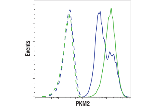
Flow cytometric analysis of 293T cells, transfected with PKM2 siRNA (blue) or mock transfected (green), using PKM2 (D78A4) XP® Rabbit mAb (solid lines) or a concentration-matched Rabbit (DA1E) mAb IgG XP® Isotype Control #3900 (dashed lines). Anti-rabbit IgG (H+L), F(ab')2 Fragment (PE Conjugate) #8885 was used as a secondary antibody.
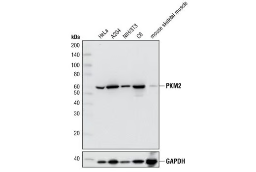
Western blot analysis of extracts from various cell lines and mouse skeletal muscle using PKM2 (D78A4) XP® Rabbit mAb (upper) or GAPDH (14C10) Rabbit mAb #2118.
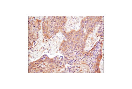
Immunohistochemical analysis of paraffin-embedded human lung carcinoma using PKM2 (D78A4) XP® Rabbit mAb.
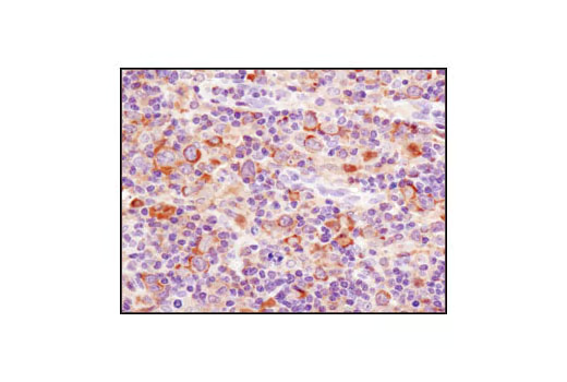
Immunohistochemical analysis of paraffin-embedded human lymphoma using PKM2 (D78A4) XP® Rabbit mAb.
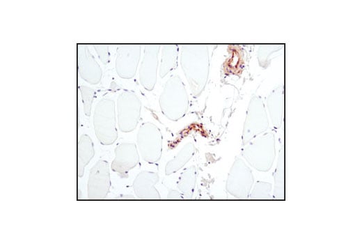
Immunohistochemical analysis of paraffin-embedded human skeletal muscle using PKM2 (D78A4) XP® Rabbit mAb. Note the lack of staining in the skeletal muscle cells which do not express PKM2 while vessels within the tissue stain positively.
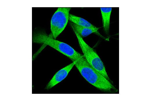
Confocal immunofluorescent analysis of A204 cells using PKM2 (D78A4) XP® Rabbit mAb (green). Blue pseudocolor = DRAQ5® #4084 (fluorescent DNA dye).