The T47-530 specifically recognizes the Lymphocyte Activation Gene 3 (LAG-3) protein which is also known as, Protein FDC, or CD223. LAG-3 is a ~70 kDa type I transmembrane glycoprotein that belongs to the Ig superfamily and exhibits homology to CD4. LAG-3 is expressed on NK cells, regulatory T cells, and activated conventional T cells with higher expression found on CD8+ T cells compared with CD4+ T cells. LAG-3 is an activation induced cell surface molecule that like CD4, binds MHC class II molecules, but with much higher affinity. This may enable LAG-3 to act as a negative competitor of CD4 for MHC class II ligand binding. LAG-3 may associate with the TCR-CD3 complex to downregulate TCR signal transduction and T cell clonal expansion. In contrast, LAG-3-induced signaling may promote dendritic cell activation.
商品描述
T47-530
The T47-530 specifically recognizes the Lymphocyte Activation Gene 3 (LAG-3) protein which is also known as, Protein FDC, or CD223. LAG-3 is a ~70 kDa type I transmembrane glycoprotein that belongs to the Ig superfamily and exhibits homology to CD4. LAG-3 is expressed on NK cells, regulatory T cells, and activated conventional T cells with higher expression found on CD8+ T cells compared with CD4+ T cells. LAG-3 is an activation induced cell surface molecule that like CD4, binds MHC class II molecules, but with much higher affinity. This may enable LAG-3 to act as a negative competitor of CD4 for MHC class II ligand binding. LAG-3 may associate with the TCR-CD3 complex to downregulate TCR signal transduction and T cell clonal expansion. In contrast, LAG-3-induced signaling may promote dendritic cell activation.
同种型
Mouse IgG1, κ
克隆号
克隆 T47-530 (RUO)
产品详情
PE
R-Phycoerythrin (PE), is part of the BD family of Phycobiliprotein dyes. This fluorochrome is a multimeric fluorescent phycobiliprotein with excitation maximum (Ex Max) of 496 nm and 566 nm and an emission maximum (Em Max) at 576 nm. PE is designed to be excited by the Blue (488 nm), Green (532 nm) and Yellow-Green (561 nm) lasers and detected using an optical filter centered near 575 nm (e.g., a 575/26-nm bandpass filter). As PE is excited by multiple lasers, this can result in cross-laser excitation and fluorescence spillover on instruments with various combinations of Blue, Green, and Yellow-Green lasers. Please ensure that your instrument’s configurations (lasers and optical filters) are appropriate for this dye.
PE
Yellow-Green 488 nm, 532 nm, 561 nm
496 nm, 566 nm
576 nm
应用
实验应用
Flow cytometry (Routinely Tested)
推荐用量
5 µl
反应种属
Human (QC Testing)
目标/特异性
CD223 (LAG-3)
背景
别名
LAG3; CD223; FDC; Lymphocyte activation gene 3 protein; Protein FDC
制备和贮存
存储溶液
Aqueous buffered solution containing BSA and ≤0.09% sodium azide.
保存方式
Aqueous buffered solution containing BSA and ≤0.09% sodium azide.
文献
文献
研发参考(5)
1. Casati C, Camisaschi C, Novellino L, et al. Human lymphocyte activation gene-3 molecules expressed by activated T cells deliver costimulation signal for dendritic cell activation. J Immunol. 2008; 180(6):3782-3788. (Biology).
2. Hannier S, Tournier M, Bismuth G, Triebel F. CD3/TCR complex-associated lymphocyte activation gene-3 molecules inhibit CD3/TCR signaling. J Immunol. 1998; 161(8):4058-4065. (Biology).
3. Huang CT, Workman CJ, Flies D, et al. Role of LAG-3 in regulatory T cells. Immunity. 2004; 21(4):503-513. (Biology).
4. Triebel F, Hacene K, Pichon MF. A soluble lymphocyte activation gene-3 (sLAG-3) protein as a prognostic factor in human breast cancer expressing estrogen or progesterone receptors. Cancer Lett. 2006; 235(1):147-153. (Biology).
5. Triebel F, Jitsukawa S, Baixeras E, et al. LAG-3, a novel lymphocyte activation gene closely related to CD4. J Exp Med. 1990; 171(5):1393-1405. (Biology).
数据库链接
Entrez-Gene ID
3902
参考图片
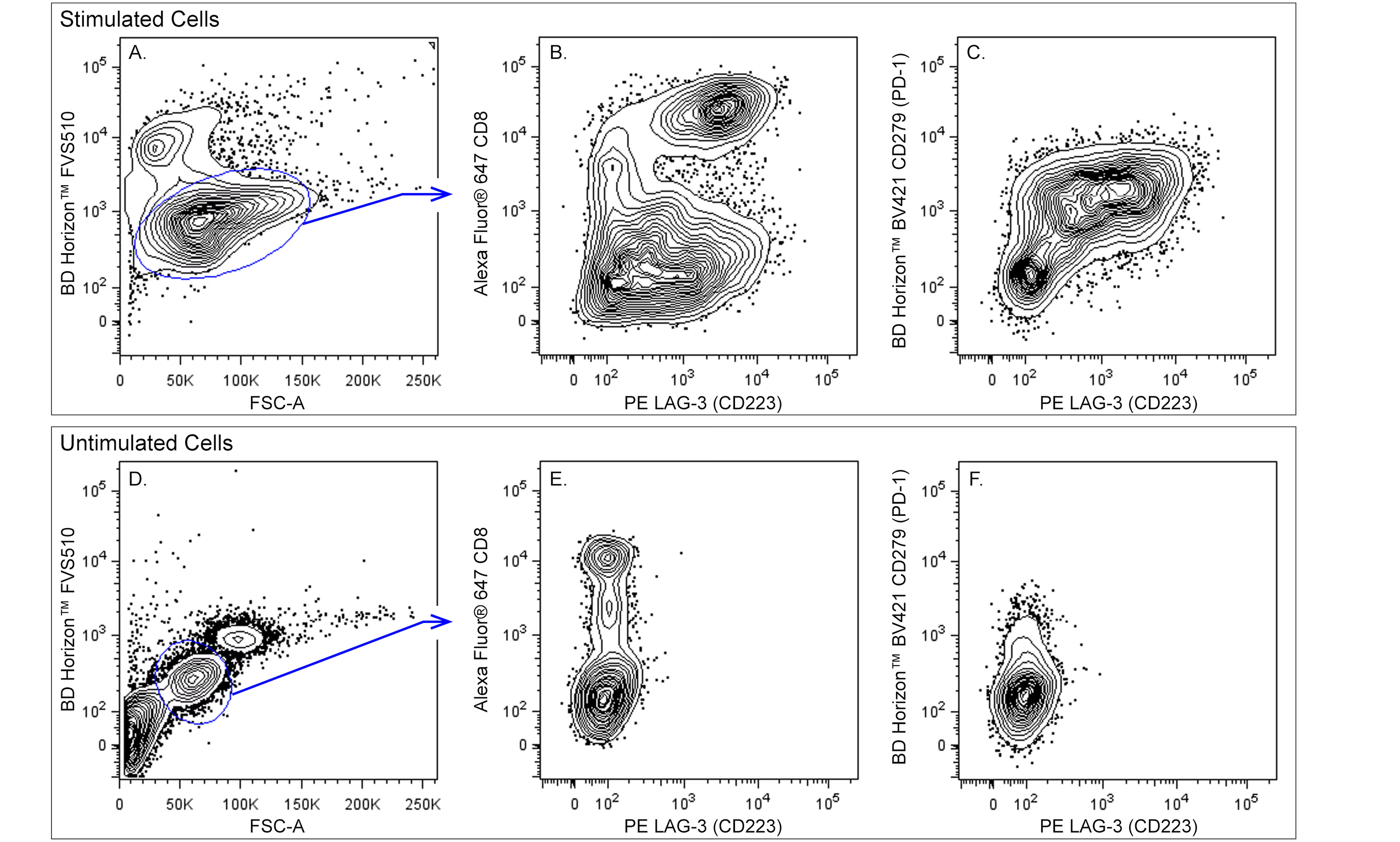
Multicolor flow cytometric analysis of LAG-3 (CD223) expression on activated human peripheral blood lymphocytes. Human peripheral blood mononuclear cells were left untreated (Unstimulated Cells; Bottom Panel), or were cultured (Stimulated Cells; Top Panel) for 3 days with plate-bound Anti-Human CD3 (Cat. No. 555329; 10 µg/mL for coating) and soluble Anti-Human CD28 (Cat. No. 555725; 1 µg/mL) antibodies, and Human Recombinant IL-2 (Cat. No. 554603; 10 ng/mL). The Unstimulated and Stimulated Cells were pre-stained with BD Horizon Fixable Viability Stain 510 (FVS510; Cat. No. 564406) and fixed with BD Cytofix™ Fixation Buffer (Cat. No. 554655). The cells were then stained with Alexa Fluor® 647 Mouse Anti-Human CD8 (Cat. No. 557708), BD Horizon™ BV421 Mouse Anti-Human CD279 (PD-1) (Cat. No. 562516), and PE Mouse Anti-Human LAG-3 (CD223) (Cat. No. 565616/ 565617) antibodies. Two-color flow cytometric contour plots showing the correlated expression of LAG-3 (CD223) versus CD8 (Plots B and E) or CD279 (PD-1) (Plots C and F) were derived from live lymphocyte-gated events [ie, gated events with low level FVS510 incorporation and forward scattered-light signals (FSC-A) characteristic of intact lymphocytes; Plots A and D]. Flow cytometric analysis was performed using a BD LSRFortessa™ Flow Cytometer System.


 全部商品分类


 用小程序,查商品更便捷


