


 全部商品分类
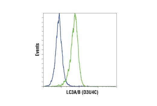


LC3A/B (D3U4C) XP ® Rabbit mAb

 下载产品说明书 下载COA 下载SDS
 用小程序,查商品更便捷


 收藏
收藏
 对比
对比 咨询
咨询
Monoclonal antibody is produced by immunizing animals with a synthetic peptide corresponding to residues surrounding Leu44 of human LC3B protein (conserved in LC3A).
Product Usage Information
| Application | Dilution |
|---|---|
| Western Blotting | 1:1000 |
| Immunohistochemistry (Paraffin) | 1:250 - 1:1000 |
| Immunofluorescence (Immunocytochemistry) | 1:50 - 1:200 |
| Flow Cytometry (Fixed/Permeabilized) | 1:100 - 1:400 |
Specificity/Sensitivity
Species Reactivity:
Human, Mouse, Rat
Supplied in 10 mM sodium HEPES (pH 7.5), 150 mM NaCl, 100 µg/ml BSA, 50% glycerol and less than 0.02% sodium azide. Store at –20°C. Do not aliquot the antibody.
For a carrier free (BSA and azide free) version of this product see product #58139.
参考图片
Flow cytometric analysis of HeLa cells, untreated (blue) or treated with chloroquine (50 µM, 16 hr; green) using LC3A/B (D3U4C) Rabbit mAb. Anti-rabbit IgG (H+L), F(ab')2 Fragment (Alexa Fluor® 647 Conjugate) #4414 was used as a secondary antibody.
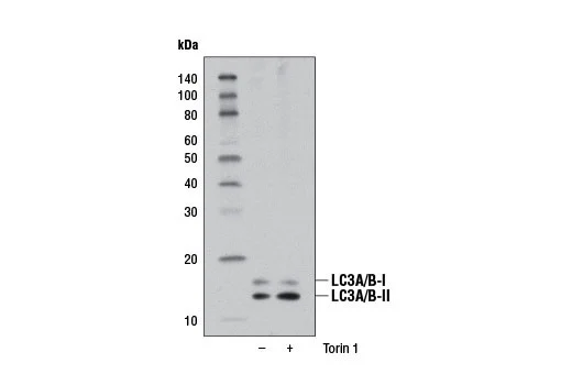
Western blot analysis of extracts from RD cells, untreated (-) or Torin 1-treated (250 nM, 4 hr; +), using LC3A/B (D3U4C) XP® Rabbit mAb.
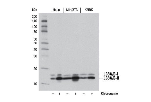
Western blot analysis of extracts from HeLa, NIH/3T3, and KNRK cells, untreated (-) or chloroquine-treated (50 μM, overnight; +), using LC3A/B (D3U4C) XP® Rabbit mAb.
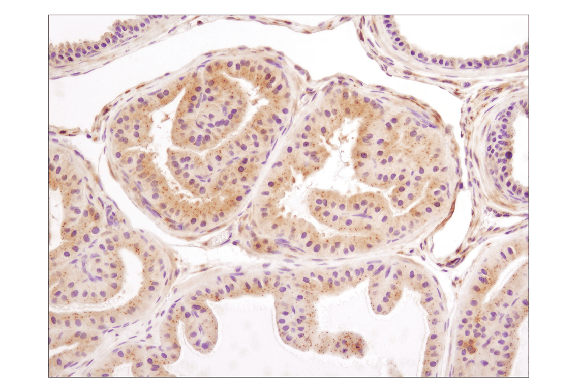
Immunohistochemical analysis of paraffin-embedded mouse prostate using LC3A/B (D3U4C) XP® Rabbit mAb.
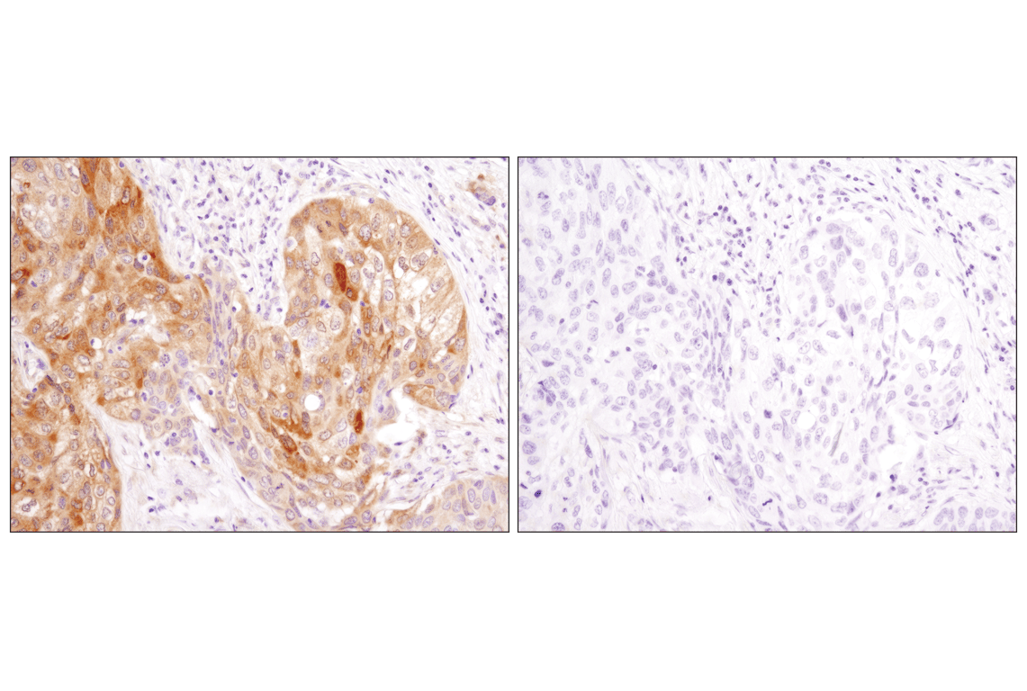
Immunohistochemical analysis of paraffin-embedded human squamous cell lung carcinoma using LC3A/B (D3U4C) XP® Rabbit mAb in the presence of control peptide (left) or antigen-specific peptide (right).
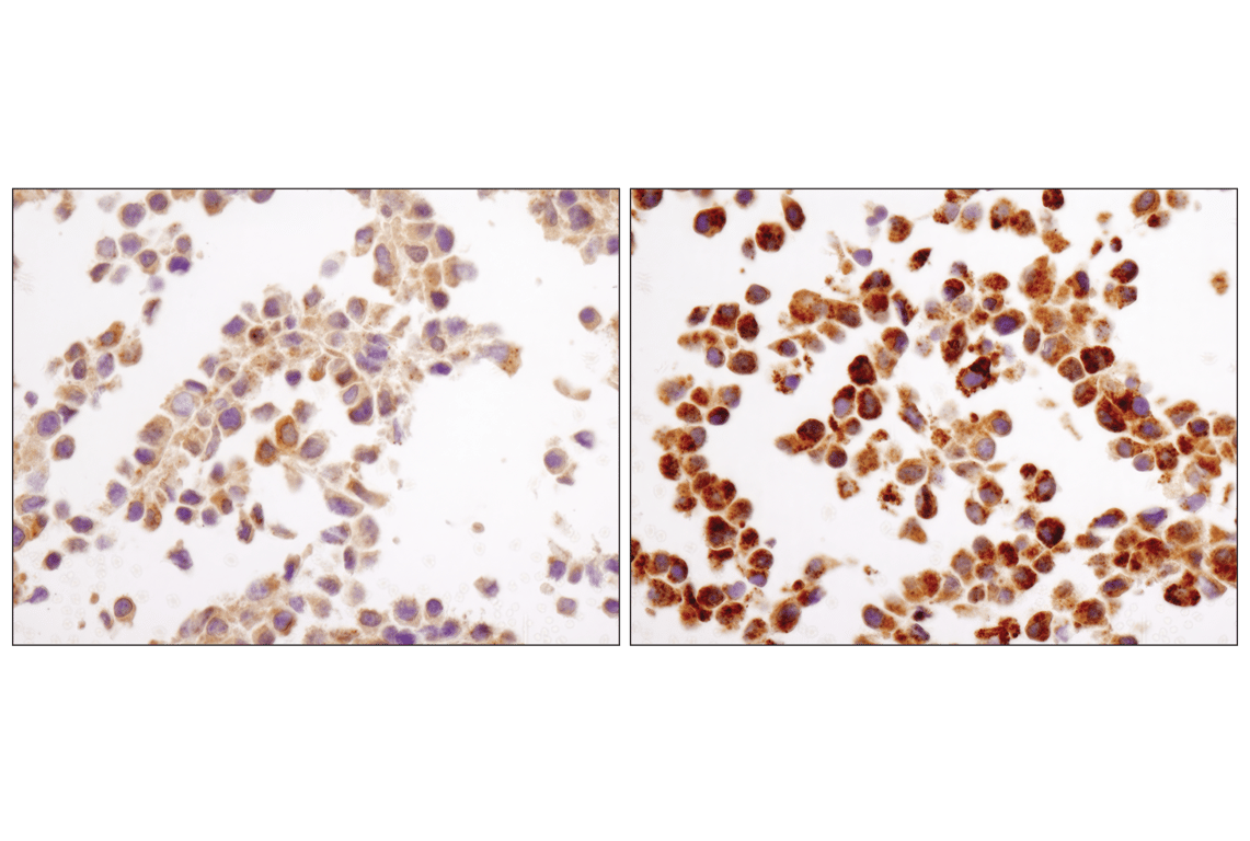
Immunohistochemical analysis of paraffin-embedded NIH/3T3 cell pellets, control (left) or chloroquine-treated (right), using LC3A/B (D3U4C) XP® Rabbit mAb.
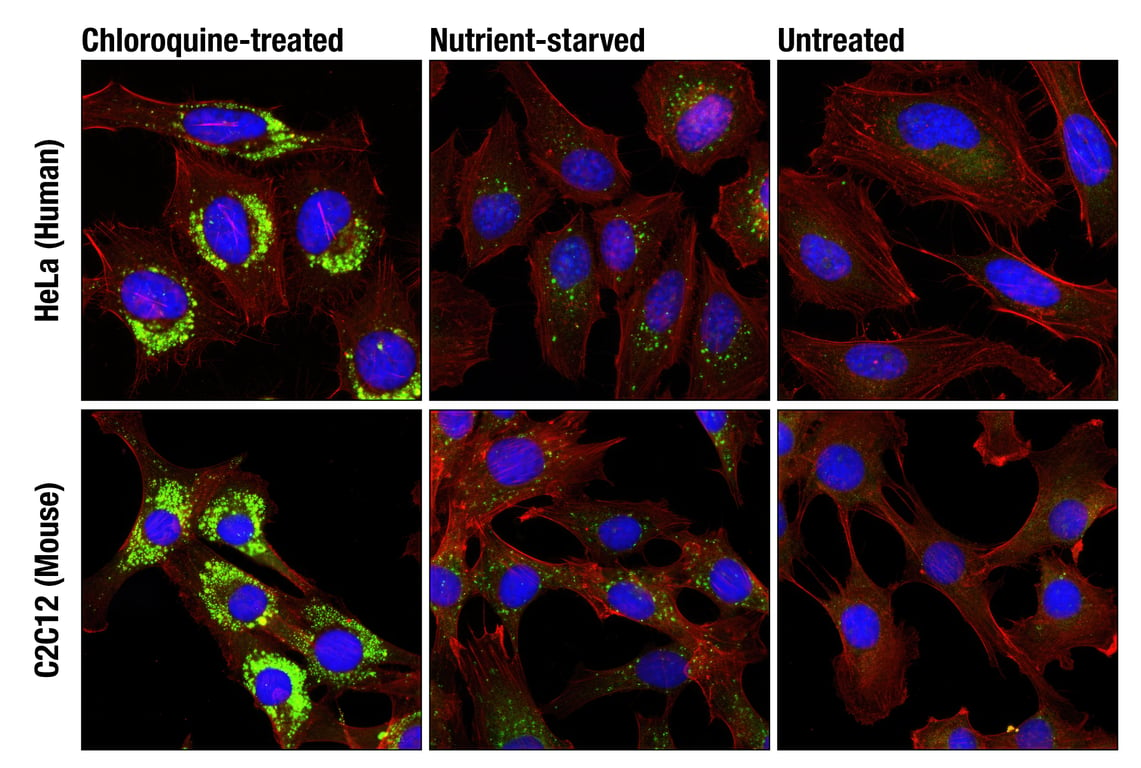
Confocal immunofluorescent analysis of HeLa (upper) and C2C12 (lower) cells, chloroquine-treated (50 μM, overnight; left), nutrient-starved with EBSS (3 hr, middle) or untreated (right) using LC3A/B (D3U4C) XP® Rabbit mAb (green) and β-Actin (13E5) Rabbit mAb (Alexa Fluor® 555 Conjugate) #8046 (red). Blue pseudocolor= DRAQ5® #4084 (fluorescent DNA dye).