


 全部商品分类


 用小程序,查商品更便捷


Polyclonal antibodies are produced by immunizing animals with a synthetic peptide corresponding to residues surrounding Gly40 of LC3B. Antibodies are purified by protein A and peptide affinity chromatography.
Product Usage Information
| Application | Dilution |
|---|---|
| Western Blotting | 1:1000 |
| Immunofluorescence (Immunocytochemistry) | 1:100 - 1:400 |
| Flow Cytometry (Fixed/Permeabilized) | 1:50 - 1:100 |
Specificity/Sensitivity
Species Reactivity:
Human, Mouse, Rat
Supplied in 10 mM sodium HEPES (pH 7.5), 150 mM NaCl, 100 µg/ml BSA and 50% glycerol. Store at –20°C. Do not aliquot the antibody.
参考图片
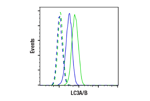
Flow cytometric analysis of ACHN cells, untreated (blue) or treated with Chloroquine #14774 (50 μM, 16 hr; green) using LC3A/B Antibody (solid lines) or concentration-matched Rabbit (DA1E) mAb IgG XP® Isotype Control #3900 (dashed lines). Anti-rabbit IgG (H+L), F(ab')2 Fragment (Alexa Fluor® 488 Conjugate) #4412 was used as a secondary antibody.
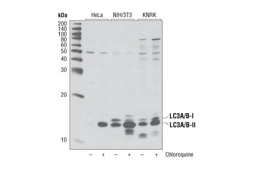
Western blot analysis of extracts from various cell lines, untreated or treated with chloroquine (50 μM, overnight) using LC3A/B Antibody.
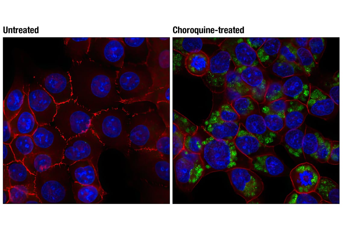
Confocal immunofluorescent analysis of HCT-116 cells, untreated (left) or choroquine-treated (50 uM, overnight; right) using LC3A/B Antibody (green) and β-Catenin (L54E2) Mouse mAb (Alexa Fluor® 555 Conjugate) #5612 (red). Blue pseudocolor = DRAQ5® #4084 (fluorescent DNA dye).