


 全部商品分类
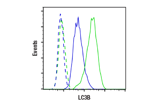


LC3B (E5Q2K) Mouse mAb

 下载产品说明书 下载COA 下载SDS
 用小程序,查商品更便捷


 收藏
收藏
 对比
对比 咨询
咨询
Monoclonal antibody is produced by immunizing animals with a synthetic peptide corresponding to residues near the amino terminus of human LC3B protein.
Product Usage Information
| Application | Dilution |
|---|---|
| Western Blotting | 1:1000 |
| Immunoprecipitation | 1:100 |
| Immunohistochemistry (Paraffin) | 1:50 - 1:200 |
| Immunofluorescence (Frozen) | 1:50 - 1:200 |
| Immunofluorescence (Immunocytochemistry) | 1:200 - 1:800 |
| Flow Cytometry (Fixed/Permeabilized) | 1:100 - 1:400 |
Specificity/Sensitivity
Species Reactivity:
Human, Mouse, Rat
Supplied in 10 mM sodium HEPES (pH 7.5), 150 mM NaCl, 100 µg/ml BSA, 50% glycerol and less than 0.02% sodium azide. Store at –20°C. Do not aliquot the antibody.
For a carrier free (BSA and azide free) version of this product see product #20371.
参考图片
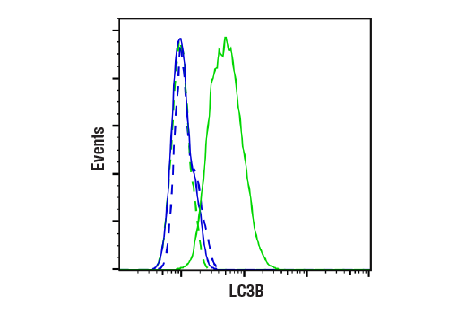
Flow cytometric analysis of HCT-116 cells, untreated (blue) or treated with Chloroquine #14774 (50 μM, 18 hr; green) using LC3B (E5Q2K) Mouse mAb Antibody (solid lines) or concentration-matched Mouse (E7Q5L) mAb IgG2b Isotype Control #53484 (dashed lines). Anti-mouse IgG (H+L), F(ab')2 Fragment (Alexa Fluor® 488 Conjugate) #4408 was used as a secondary antibody.
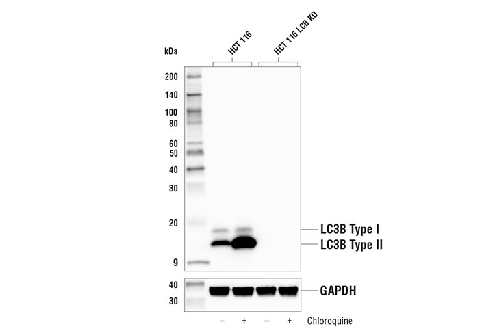
Western blot analysis of extracts from HCT 116 and HCT 116 LC3B knockout cells, untreated (-) or treated with Chloroquine #14774 (50 μM, 18 hr) using LC3B (E5Q2K) Mouse mAb #83506 (upper) or GAPDH (D16H11) XP® Rabbit mAb #5174 (lower). The absence of signal in the HCT 116 knockout cells confirms the specificity of the antibody for LC3B.
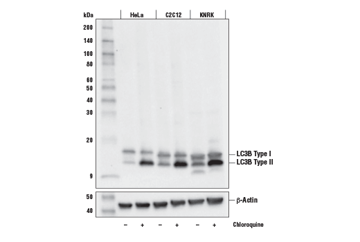
Western blot analysis of extracts from HeLa, C2C12, and KNRK cells, untreated (-) or treated with Chloroquine (50 μM, overnight; +) #14774 using LC3B (E5Q2K) Mouse mAb (upper) or β-Actin (D6A8) Rabbit mAb #8457 (lower).
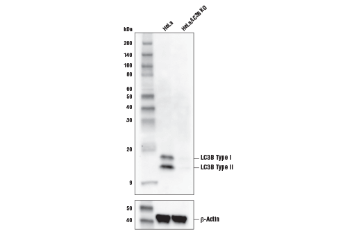
Western blot analysis of extracts from HeLa cells or HeLa cells with a knockout of LC3B (HeLa/LC3B KO) using LC3B (E5Q2K) Mouse mAb (upper) or β-Actin (D6A8) Rabbit mAb #8457 (lower).
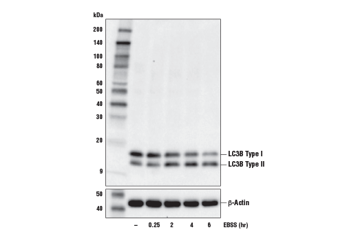
Western blot analysis of extracts from A549 cells, untreated (-) or starved with Earle's Balanced Salt Solution (EBSS) (indicated times) using LC3B (E5Q2K) Mouse mAb (upper) or β-Actin (D6A8) Rabbit mAb (lower).
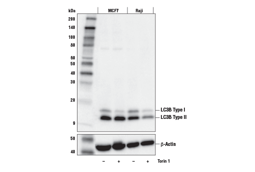
Western blot analysis of extracts from MCF7 and Raji cells, untreated (-) or treated with Torin 1 (250 nM, 5 hr; +) #14379, using LC3B (E5Q2K) Mouse mAb (upper) or β-Actin (D6A8) Rabbit mAb #8457 (lower).
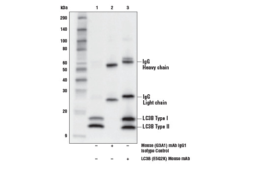
Immunoprecipitation of LC3B from HeLa cells treated with Chloroquine (50 μM, overnight) # 14774. Lane 1 is 10% input, lane 2 is Mouse (G3A1) mAb IgG1 Isotype Control, and lane 3 is LC3B (E5Q2K) Mouse mAb. Western blot was performed using LC3B (E5Q2K) Mouse mAb. Anti-mouse IgG, HRP-linked Antibody #7076 was used as a secondary antibody.
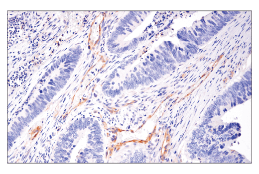
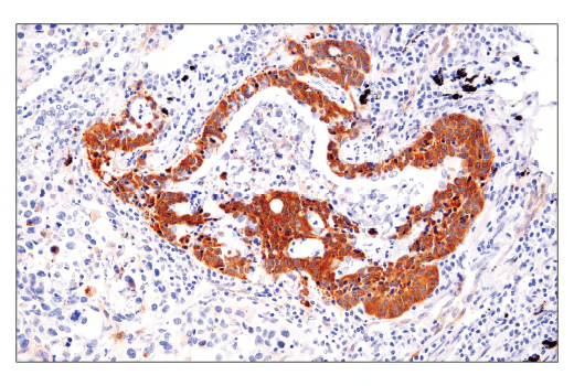
Immunohistochemical analysis of paraffin-embedded human colon carcinoma using LC3B (E5Q2K) Mouse mAb.
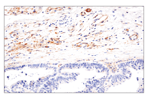
Immunohistochemical analysis of paraffin-embedded human esophageal carcinoma using LC3B (E5Q2K) Mouse mAb.
Immunohistochemical analysis of paraffin-embedded human lung carcinoma using LC3B (E5Q2K) Mouse mAb.
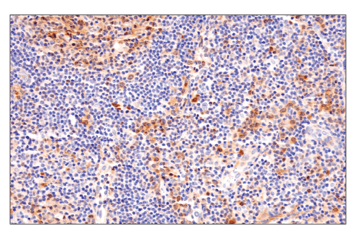
Immunohistochemical analysis of paraffin-embedded human non-Hodgkin's lymphoma using LC3B (E5Q2K) Mouse mAb.
Immunohistochemical analysis of paraffin-embedded HCT116 cell pellets, untreated (left-top) or treated with Chloroquine #14774 (right-top), and HCT116 LC3B knockout cell pellets, untreated (left-bottom) or treated with Chloroquine #14774 (right-bottom), using LC3B (E5Q2K) Mouse mAb.
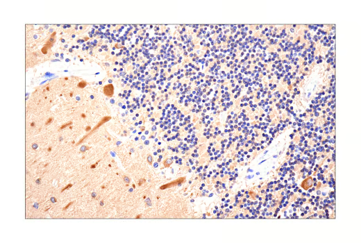
Immunohistochemical analysis of paraffin-embedded normal human brain using LC3B (E5Q2K) Mouse mAb.
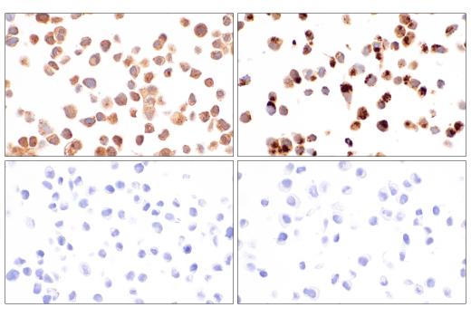
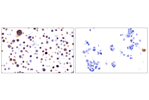
Immunohistochemical analysis of paraffin-embedded KARPAS 299 cell pellet (left, high-expressing) or VCaP cell pellet (right, low-expressing) using LC3B (E5Q2K) Mouse mAb.
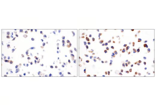
Immunohistochemical analysis of paraffin-embedded HeLa cell pellets, untreated (left) or treated with Chloroquine #14774 (right), using LC3B (E5Q2K) Mouse mAb.
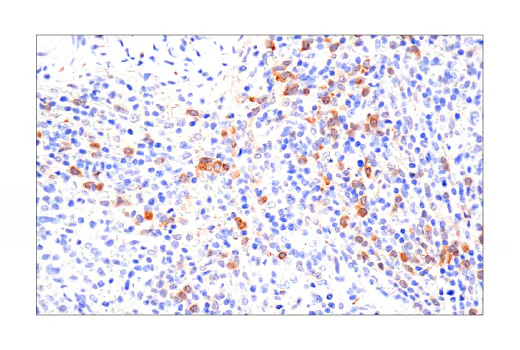
Immunohistochemical analysis of paraffin-embedded normal human spleen using LC3B (E5Q2K) Mouse mAb.
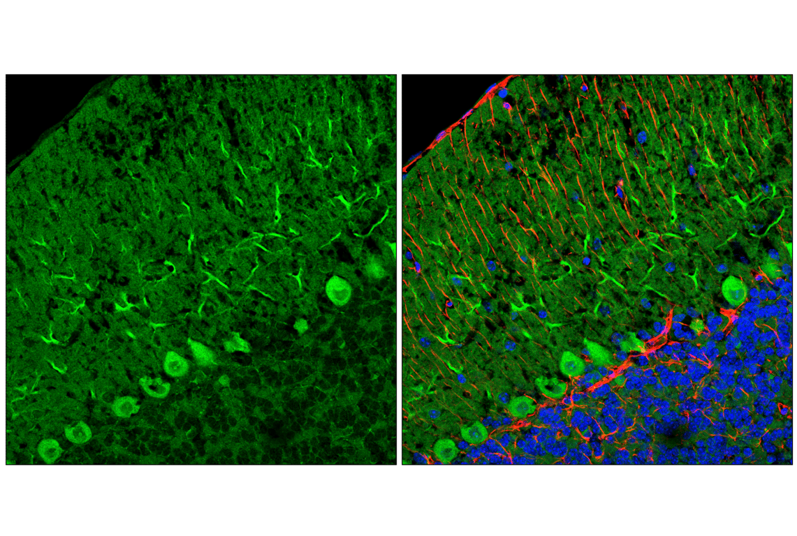
Confocal immunofluorescent analysis of fixed frozen mouse cerebellum, labeled with LC3B (E5Q2K) Mouse mAb #83506 (left, green) and co-labeled with GFAP (E4L7M) XP® Rabbit mAb #80788 (right, red) and DAPI #4083 (right, blue).
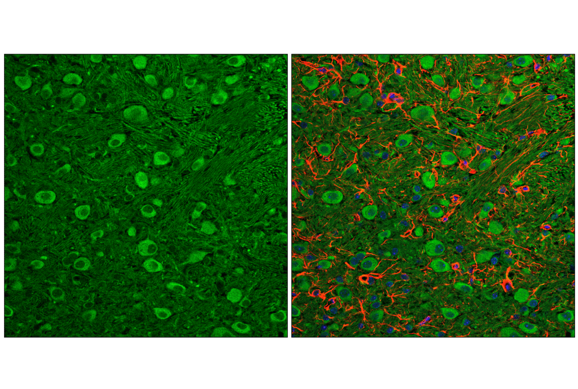
Confocal immunofluorescent analysis of fixed frozen mouse pons, labeled with LC3B (E5Q2K) Mouse mAb #83506 (left, green) and co-labeled with GFAP (E4L7M) XP® Rabbit mAb #80788 (right, red) and DAPI #4083 (right, blue).
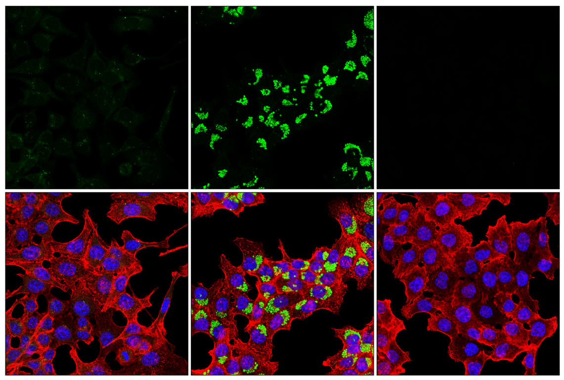
Confocal immunofluorescent analysis of HCT 116 cells either untreated (left) or treated with Chloroquine #14774 (50 µM, overnight) (center) or LC3B HCT 116 knockout cells treated with Chloroquine #14774 (50 µM, overnight) (right) using LC3B (E5Q2K) Mouse mAb (green). Actin filaments were labeled with β-Actin (13E5) Rabbit mAb #4970 (red) and nuclei were labeled with DAPI #4083 (blue).
Flow cytometric analysis of HCT-116 cells, wild-type (green, high expression) or LC3B knockdown (blue, negative expression), using LC3B (E5Q2K) Mouse mAb or a concentration-matched Mouse (E7Q5L) mAb IgG2b Isotype Control #53484 (dashed lines). Anti-mouse IgG (H+L), F(ab')2 Fragment (Alexa Fluor® 488 Conjugate) #4408 was used as a secondary antibody.