




 全部商品分类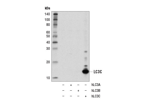


1/2


 用小程序,查商品更便捷





产品介绍
产品介绍
产品信息
荧光素标记
Unconjugated

抗原名称
LC3C
来源纯化
Monoclonal antibody is produced by immunizing animals with a synthetic peptide corresponding to residues surrounding Leu50 of human LC3C protein.
宿主
Rabbit
商品描述
Product Usage Information
| Application | Dilution |
|---|---|
| Western Blotting | 1:1000 |
同种型
Rabbit IgG
分子量
14
研究领域
癌症,细胞生物学,纤维化,代谢,神经科学
应用
反应种属
Human
目标/特异性
Specificity/Sensitivity
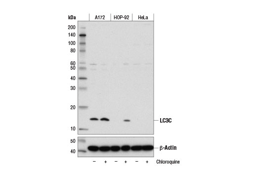
LC3C (D1R8V) Rabbit mAb recognizes endogenous levels of total LC3C protein. This antibody does not cross react with other LC3 isoforms.
Species Reactivity:
Human
敏感性
Endogenous
背景
背景
Autophagy is a catabolic process for the autophagosomic-lysosomal degradation of bulk cytoplasmic contents (1,2). Autophagy is generally activated by conditions of nutrient deprivation, but it has also been associated with a number of physiological processes including development, differentiation, neurodegenerative diseases, infection, and cancer (3). Autophagy marker Light Chain 3 (LC3) was originally identified as a subunit of microtubule-associated proteins 1A and 1B (termed MAP1LC3) (4) and subsequently found to contain similarity to the yeast protein Apg8/Aut7/Cvt5 critical for autophagy (5). Three human LC3 isoforms (LC3A, LC3B, and LC3C) undergo posttranslational modifications during autophagy (6-9). Cleavage of LC3 at the carboxy terminus immediately following synthesis yields the cytosolic LC3-I form. During autophagy, LC3-I is converted to LC3-II through lipidation by a ubiquitin-like system involving Atg7 and Atg3 that allows for LC3 to become associated with autophagic vesicles (6-10). The presence of LC3 in autophagosomes and the conversion of LC3 to the lower migrating form, LC3-II, have been used as indicators of autophagy (11).In general, expression of LC3C protein is lower than the other LC3 isoforms, with the highest LC3C expression observed in placenta, lung, and ovary (7).
1.Reggiori, F. and Klionsky, D.J. (2002) Eukaryot. Cell 1, 11-21.
2.Codogno, P. and Meijer, A.J. (2005) Cell Death Differ. 12 Suppl 2, 1509-18.
3.Levine, B. and Yuan, J. (2005) J. Clin. Invest. 115, 2679-88.
4.Mann, S.S. and Hammarback, J.A. (1994) J. Biol. Chem. 269, 11492-97.
5.Lang, T. et al. (1998) EMBO J. 17, 3597-607.
6.Kabeya, Y. et al. (2000) EMBO J. 19, 5720-28.
7.He, H. et al. (2003) J. Biol. Chem. 278, 29278-87.
8.Tanida, I. et al. (2004) J. Biol. Chem. 279, 47704-10.
9.Wu, J. et al. (2006) Biochem. Biophys. Res. Commun. 339, 437-42.
10.Ichimura, Y. et al. (2000) Nature 408, 488-92.
11.Kabeya, Y. et al. (2004) J. Cell Sci. 117, 2805-12.
研究领域
癌症,细胞生物学,纤维化,代谢,神经科学
翻译后修饰
unmodified
制备和贮存
保存方式
Supplied in 10 mM sodium HEPES (pH 7.5), 150 mM NaCl, 100 µg/ml BSA, 50% glycerol and less than 0.02% sodium azide. Store at –20°C. Do not aliquot the antibody.
数据库链接
Entrez-Gene ID
440738
UniProt ID
Q9BXW4
声明 :本官网所有报价均为常温或者蓝冰运输价格,如有产品需要干冰运输,需另外加收干冰运输费。