


 全部商品分类
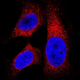


hLDHB MAb (Cl 2057D) (25 ug)

 下载产品说明书 下载SDS
 用小程序,查商品更便捷


 收藏
收藏
 对比
对比 咨询
咨询
Met1-Val93
Accession # P07195
Scientific Data
 View Larger
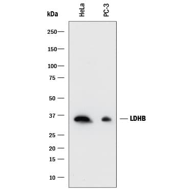
Detection of Human Lactate Dehydrogenase B/LDHB by Western Blot. Western blot shows lysates of HeLa human cervical epithelial carcinoma cell line and PC-3 human prostate cancer cell line. PVDF membrane was probed with 0.05 µg/mL of Rabbit Anti-Human Lactate Dehydrogenase B/LDHB Monoclonal Antibody (Catalog # MAB9205) followed by HRP-conjugated Anti-Rabbit IgG Secondary Antibody (Catalog # HAF008). A specific band was detected for Lactate Dehydrogenase B/LDHB at approximately 36 kDa (as indicated). This experiment was conducted under reducing conditions and using Immunoblot Buffer Group 1.
.") View Larger
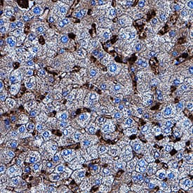
View LargerLactate Dehydrogenase B/LDHB in Human Liver. Lactate Dehydrogenase B/LDHB was detected in immersion fixed paraffin-embedded sections of human liver using Rabbit Anti-Human Lactate Dehydrogenase B/LDHB Monoclonal Antibody (Catalog # MAB9205) at 1 µg/mL for 1 hour at room temperature followed by incubation with the Anti-Rabbit IgG VisUCyte™ HRP Polymer Antibody (Catalog # VC003). Tissue was stained using DAB (brown) and counterstained with hematoxylin (blue). Specific staining was localized to sinusoids. View our protocol for IHC Staining with VisUCyte HRP Polymer Detection Reagents.
.") View Larger
View LargerLactate Dehydrogenase B/LDHB in HeLa Human Cell Line. Lactate Dehydrogenase B/LDHB was detected in immersion fixed HeLa human cervical epithelial carcinoma cell line using Rabbit Anti-Human Lactate Dehydrogenase B/LDHB Monoclonal Antibody (Catalog # MAB9205) at 3 µg/mL for 3 hours at room temperature. Cells were stained using the NorthernLights™ 557-conjugated Anti-Rabbit IgG Secondary Antibody (red; Catalog # NL004) and counterstained with DAPI (blue). Specific staining was localized to cytoplasm. View our protocol for Fluorescent ICC Staining of Cells on Coverslips.
 View Larger
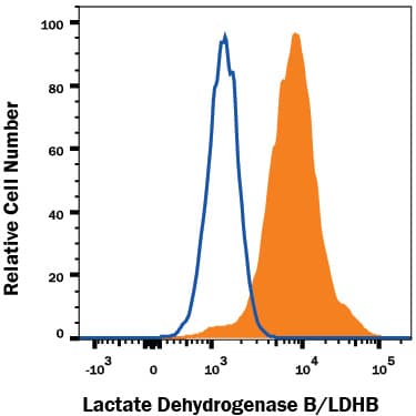
Detection of Lactate Dehydrogenase B/LDHB in HeLa Human Cell Line by Flow Cytometry. HeLa human cervical epithelial carcinoma cell line was stained with Rabbit Anti-Human Lactate Dehydrogenase B/LDHB Monoclonal Antibody (Catalog # MAB9205, filled histogram) or isotype control antibody (Catalog # AB-105-C, open histogram), followed by Allophycocyanin-conjugated Anti-Rabbit IgG Secondary Antibody (Catalog # F0111). To facilitate intracellular staining, cells were fixed with Flow Cytometry Fixation Buffer (Catalog # FC004) and permeabilized with Flow Cytometry Permeabilization/Wash Buffer I (Catalog # FC005). View our protocol for Staining Intracellular Molecules.
 View Larger
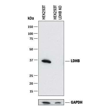
Western Blot Shows Human Lactate Dehydrogenase B/LDHB Specificity by Using Knockout Cell Line. Western blot shows lysates of HEK293T human embryonic kidney parental cell line and LDHB knockout HEK293T cell line (KO). PVDF membrane was probed with 0.05 µg/mL of Rabbit Anti-Human Lactate Dehydrogenase B/LDHB Monoclonal Antibody (Catalog # MAB9205) followed by HRP-conjugated Anti-Rabbit IgG Secondary Antibody (Catalog # HAF008). A specific band was detected for Lactate Dehydrogenase B/LDHB at approximately 36 kDa (as indicated) in the parental HEK293T cell line, but is not detectable in knockout HEK293T cell line. GAPDH (Catalog # MAB5718) is shown as a loading control. This experiment was conducted under reducing conditions and using Immunoblot Buffer Group 1.
Human Lactate Dehydrogenase B/LDHB Antibody Summary
Met1-Val93
Accession # P07195
Applications
Please Note: Optimal dilutions should be determined by each laboratory for each application. General Protocols are available in the Technical Information section on our website.
Background: Lactate Dehydrogenase B/LDHB
LDHB is one of two subunits of Lactose Dehydrogenase which is formed from tetramer arrangements of the A and B subunits. LDHB is part of the pyruvate fermentation to lactate pathway in which it catalyzes the conversion of L-lactate and NAD to pyruvate and NADH. LDHA is widely expressed, with high expression in muscle and brain. High expression in tumor cells correlates with low survival rates in various cancers including osteosarcoma, oral squamous cell carcinoma breast and lung cancers. Unlike Lactose Dehyrogenase A, LDHB mutations are not linked to any disease
Preparation and Storage
- 12 months from date of receipt, -20 to -70 °C as supplied.
- 1 month, 2 to 8 °C under sterile conditions after reconstitution.
- 6 months, -20 to -70 °C under sterile conditions after reconstitution.
参考图片
Detection of Human Lactate Dehydrogenase B/LDHB by Western Blot. Western blot shows lysates of HeLa human cervical epithelial carcinoma cell line and PC‑3 human prostate cancer cell line. PVDF membrane was probed with 0.05 µg/mL of Rabbit Anti-Human Lactate Dehydrogenase B/LDHB Monoclonal Antibody (Catalog # MAB9205) followed by HRP-conjugated Anti-Rabbit IgG Secondary Antibody (Catalog # HAF008). A specific band was detected for Lactate Dehydrogenase B/LDHB at approximately 36 kDa (as indicated). This experiment was conducted under reducing conditions and using Immunoblot Buffer Group 1.
Lactate Dehydrogenase B/LDHB in HeLa Human Cell Line. Lactate Dehydrogenase B/LDHB was detected in immersion fixed HeLa human cervical epithelial carcinoma cell line using Rabbit Anti-Human Lactate Dehydrogenase B/LDHB Monoclonal Antibody (Catalog # MAB9205) at 3 µg/mL for 3 hours at room temperature. Cells were stained using the NorthernLights™ 557-conjugated Anti-Rabbit IgG Secondary Antibody (red; Catalog # NL004) and counterstained with DAPI (blue). Specific staining was localized to cytoplasm. View our protocol for Fluorescent ICC Staining of Cells on Coverslips.
Lactate Dehydrogenase B/LDHB in Human Liver. Lactate Dehydrogenase B/LDHB was detected in immersion fixed paraffin-embedded sections of human liver using Rabbit Anti-Human Lactate Dehydrogenase B/LDHB Monoclonal Antibody (Catalog # MAB9205) at 1 µg/mL for 1 hour at room temperature followed by incubation with the Anti-Rabbit IgG VisUCyte™ HRP Polymer Antibody (Catalog # VC003). Tissue was stained using DAB (brown) and counterstained with hematoxylin (blue). Specific staining was localized to sinusoids. View our protocol for IHC Staining with VisUCyte HRP Polymer Detection Reagents.
Detection of Lactate Dehydrogenase B/LDHB in HeLa Human Cell Line by Flow Cytometry. HeLa human cervical epithelial carcinoma cell line was stained with Rabbit Anti-Human Lactate Dehydrogenase B/LDHB Monoclonal Antibody (Catalog # MAB9205, filled histogram) or isotype control antibody (Catalog # AB-105-C, open histogram), followed by Allophycocyanin-conjugated Anti-Rabbit IgG Secondary Antibody (Catalog # F0111). To facilitate intracellular staining, cells were fixed with Flow Cytometry Fixation Buffer (Catalog # FC004) and permeabilized with Flow Cytometry Permeabilization/Wash Buffer I (Catalog # FC005). View our protocol for Staining Intracellular Molecules.