


 全部商品分类


BD Horizon™ BV421 Rat Anti-Mouse Ly-6C


 下载产品说明书 下载SDS
 用小程序,查商品更便捷


 收藏
收藏
 对比
对比 咨询
咨询

参考图片
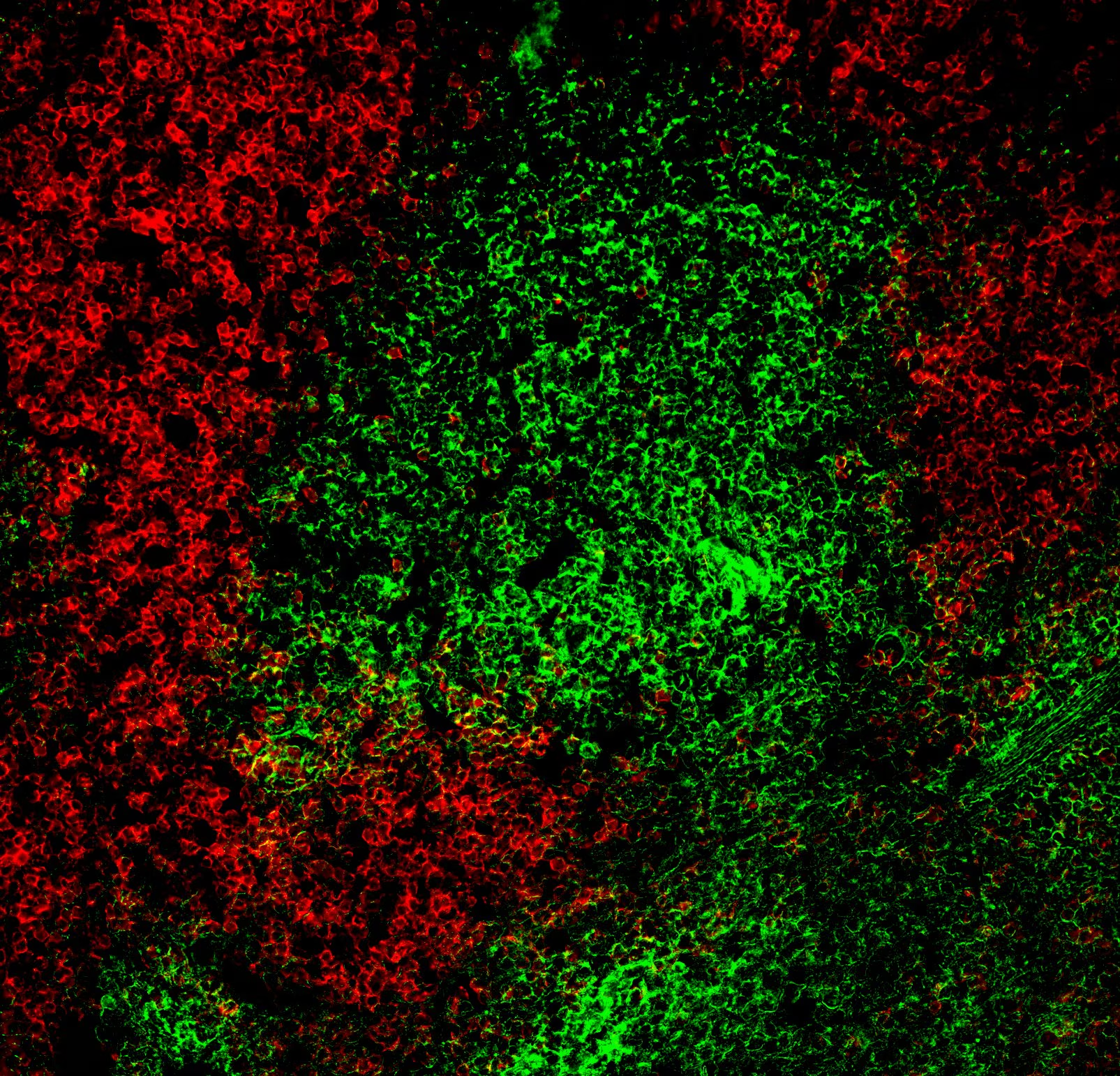
Immunohistofluorescent analysis of Ly-6C expression by cells within C57BL/6 mouse spleen. A mouse spleen cryosection (5 µm) was fixed with BD Cytofix™ Fixation Buffer (Cat. No. 554655), blocked with 5% goat serum and 1% BSA diluted in 1x PBS, and stained with BD Horizon™ BV421 Rat Anti-Mouse Ly-6C antibody (Cat. No. 562727, pseudo-colored green) and Alexa Fluor® 488 Rat Anti-Mouse CD45R/B220 antibody (Cat. No. 557669, pseudo-colored red). Images were captured on a standard epifluorescence microscope. Original magnification, 20x.
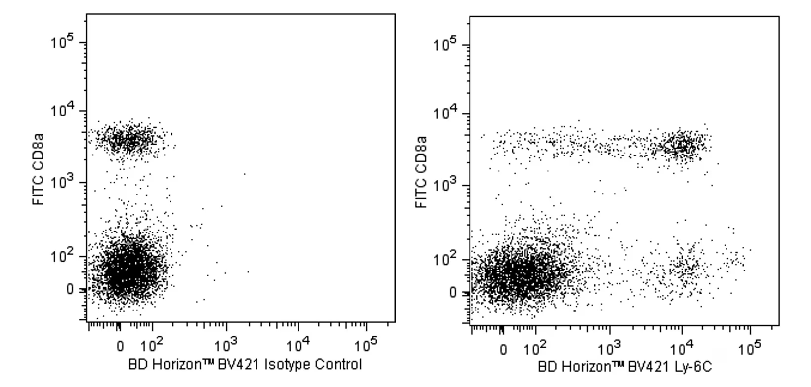
Multicolor flow cytometric analysis of Ly-6C expression on mouse splenocytes. Splenocytes from a BALB/c mouse were stained with FITC Rat Anti-Mouse CD8a antibody (Cat. No. 553031/553030/561966) and with either BD Horizon™ BV421 Rat IgM, κ Isotype Control (Cat. No. 562708, Left Panel) or with BD Horizon™ BV421 Rat Anti-Mouse Ly-6C antibody (Cat. No. 562727, Right Panel). Two-color flow cytometric dot plots showing the correlated expression of Ly-6C (or Ig isotype control staining) versus CD8a were derived from gated events with the forward and side light-scattering characteristics of viable splenocytes. Flow cytometry was performed with a BD™ LSR II Flow Cytometry System.
Immunohistofluorescent analysis of Ly-6C expression by cells within C57BL/6 mouse spleen. A mouse spleen cryosection (5 µm) was fixed with BD Cytofix™ Fixation Buffer (Cat. No. 554655), blocked with 5% goat serum and 1% BSA diluted in 1x PBS, and stained with BD Horizon™ BV421 Rat Anti-Mouse Ly-6C antibody (Cat. No. 562727, pseudo-colored green) and Alexa Fluor® 488 Rat Anti-Mouse CD45R/B220 antibody (Cat. No. 557669, pseudo-colored red). Images were captured on a standard epifluorescence microscope. Original magnification, 20x.