


 全部商品分类


BD Pharmingen™ Purified Rat Anti-Mouse Ly-6G


 下载产品说明书 下载SDS
 用小程序,查商品更便捷


 收藏
收藏
 对比
对比 咨询
咨询
参考图片
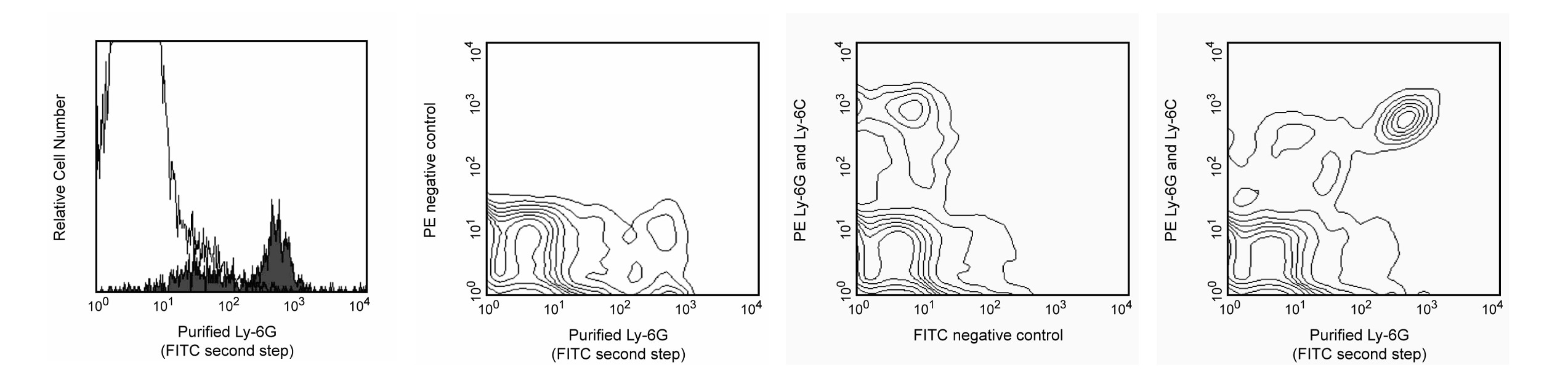
Expression of Ly-6G on peripheral-blood leukocytes. C57BL/6 whole blood was stained with purified 1A8 mAb in the presence of Mouse Fc Blockª (purified anti-mouse CD16/CD32, Cat. No. 553141/553142), followed by FITC-conjugated anti-rat IgG2a mAb RG7/1.30 (Cat. No. 553896, Far and middle left panels, right panel), then PE-conjugated anti-mouse Ly-6G and Ly-6C mAb RB6-8C5 (Cat. No. 553128, far and middle right panels). Erythrocytes were lysed (PharmLyse™, Cat. No. 555899), non-viable leukocytes were excluded by staining with propidium iodide, and leukocyte subsets were distinguished by their light-scatter profiles. Far left panel displays the expression of Ly-6G on granulocytes filled histogram) and lymphocytes/monocytes (open histogram). Note that Ly-6G expression is almost exclusively on granulocytes. Middle left and far and middle right panels compare the staining patterns of mAbs 1A8 and RB6-8C5 on total blood leukocytes. It is evident that mAb 1A8 stains the RB6-8C5-bright population, corresponding to Ly-6G-expressing granulocytes; whereas, the RB6-8C5-dim population is 1A8-negative and corresponds to Ly-6C-expressing lymphocytes and monocytes. Flow cytometry was performed on a FACSCalibur™ (BDIS, San Jose, CA).
Expression of Ly-6G on peripheral-blood leukocytes. C57BL/6 whole blood was stained with purified 1A8 mAb in the presence of Mouse Fc Blockª (purified anti-mouse CD16/CD32, Cat. No. 553141/553142), followed by FITC-conjugated anti-rat IgG2a mAb RG7/1.30 (Cat. No. 553896, Far and middle left panels, right panel), then PE-conjugated anti-mouse Ly-6G and Ly-6C mAb RB6-8C5 (Cat. No. 553128, far and middle right panels). Erythrocytes were lysed (PharmLyse™, Cat. No. 555899), non-viable leukocytes were excluded by staining with propidium iodide, and leukocyte subsets were distinguished by their light-scatter profiles. Far left panel displays the expression of Ly-6G on granulocytes filled histogram) and lymphocytes/monocytes (open histogram). Note that Ly-6G expression is almost exclusively on granulocytes. Middle left and far and middle right panels compare the staining patterns of mAbs 1A8 and RB6-8C5 on total blood leukocytes. It is evident that mAb 1A8 stains the RB6-8C5-bright population, corresponding to Ly-6G-expressing granulocytes; whereas, the RB6-8C5-dim population is 1A8-negative and corresponds to Ly-6C-expressing lymphocytes and monocytes. Flow cytometry was performed on a FACSCalibur™ (BDIS, San Jose, CA).