BD Phosflow™ Lyse/Fix Buffer is intended to be used for the lysing and fixing of whole blood for use with intracellular flow cytometry. For example, after cell stimulation with kinase activators, such as phorbol esters, the BD Phosflow™ Lyse/Fix buffer can be used for lysing red blood cells and for fixing leukocytes in one step. This allows investigators to rapidly inactivate kinases, phosphatases and/or proteases, so that the in vivo phosphorylation state of the cell can be examined. The BD Phosflow™ Lyse/Fix Buffer has been reported to preserve the light scattering properties of the cell and may be used on human and mouse whole blood. Whole blood lysis has been shown to be as effective as density gradient centrifugation in the preparation of PBMCs for lymphocyte subset analysis. In many cases, phospho-specific antibodies may be used simultaneously with the staining of cell surface markers (e.g CD markers). Not all cell surface markers, however, are compatible with the BD Phosflow™ Lyse/Fix Buffer. For a current listing of compatible cell surface markers for use with BD Phosflow™ Lyse/Fix Buffer, investigators are encouraged to visit the following website: http://www.bdbiosciences.com/documents/antibodies_human_cellsurface_marker.pdf.
Aqueous buffered solution containing formaldehyde and proprietary ingredients.
文献
文献
Development References(3)
1. Ashmore LM, Shopp GM, Edwards BS. Lymphocyte subset analysis by flow cytometry. Comparison of three different staining techniques and effects of blood storage. J Immunol Methods. 1989; 118(2):209-215. (Biology).
2. De Paoli P, Reitano M, Battistin S, Castiglia C, Santini G. Enumeration of human lymphocyte subsets by monoclonal antibodies and flow cytometry: a comparative study using whole blood or mononuclear cells separated by density gradient centrifugation. J Immunol Methods. 1984; 72(2):349-353. (Biology).
3. Renzi P, Ginns LC. Analysis of T cell subsets in normal adults. Comparison of whole blood lysis technique to Ficoll-Hypaque separation by flow cytometry. J Immunol Methods. 1987; 98(1):53-56. (Biology).
参考图片
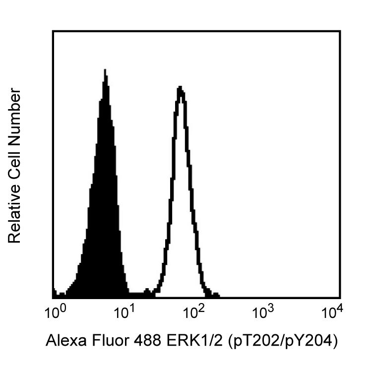
Flow cytometric analysis of ERK1/2 (pT202/pY204) on human whole blood treated with BD Phosflow™ Lyse/Fix Buffer. Freshly isolated human whole blood was left either untreated (shaded histogram) or treated with 400 nM PMA at 37°C for 10 minutes (open histogram), then fixed with Lyse/Fix Buffer 5X (Cat. No. 558049). Leukocytes were spun down and washed once with Hank's solution, followed by permeabilization with BD Phosflow™ Perm Buffer II (Cat. No. 558052) for 30 minutes. After washing the cells twice with BD Pharmingen™ Stain Buffer (Cat. No. 554656554657), the cells were stained with Alexa Fluor® 488 mouse anti-ERK1/2 (pT202/pY204) (Cat. No. 612592). Fluorescence histograms depicting ERK1/2 (pT202/pY204) expression were derived from gated events with the forward and side light-scatter characteristics of viable lymphocytes.
Flow cytometric analysis of ERK1/2 (pT202/pY204) on human whole blood treated with BD Phosflow™ Lyse/Fix Buffer. Freshly isolated human whole blood was left either untreated (shaded histogram) or treated with 400 nM PMA at 37°C for 10 minutes (open histogram), then fixed with Lyse/Fix Buffer 5X (Cat. No. 558049). Leukocytes were spun down and washed once with Hank's solution, followed by permeabilization with BD Phosflow™ Perm Buffer II (Cat. No. 558052) for 30 minutes. After washing the cells twice with BD Pharmingen™ Stain Buffer (Cat. No. 554656554657), the cells were stained with Alexa Fluor® 488 mouse anti-ERK1/2 (pT202/pY204) (Cat. No. 612592). Fluorescence histograms depicting ERK1/2 (pT202/pY204) expression were derived from gated events with the forward and side light-scatter characteristics of viable lymphocytes.


 全部商品分类


 下载产品说明书
 用小程序,查商品更便捷


 收藏
收藏
 对比
对比 咨询
咨询