


 下载产品说明书
 用小程序,查商品更便捷


 收藏
收藏
 对比
对比 咨询
咨询
Specificity/Sensitivity
参考图片
Western blot analysis of extracts from various cell lines using p38 MAPK (D13E1) XP® Rabbit mAb.
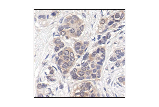
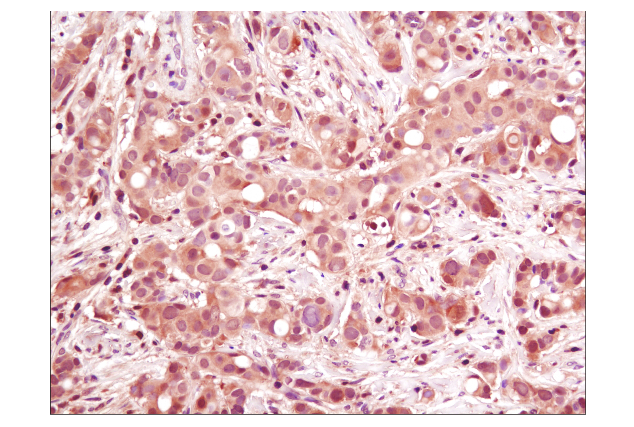
Immunohistochemical analysis of paraffin-embedded human breast carcinoma, showing cytoplasmic and nuclear localization, using p44/42 MAPK (Erk1/2) (137F5) Rabbit mAb.
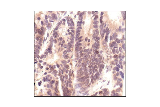
Immunohistochemical analysis of paraffin-embedded human colon carcinoma, using p44/42 MAPK (Erk1/2) (137F5) Rabbit mAb.
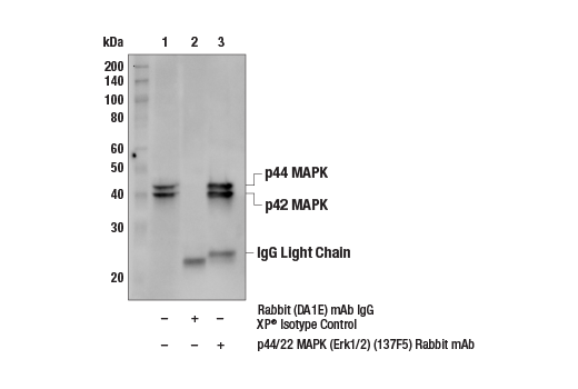
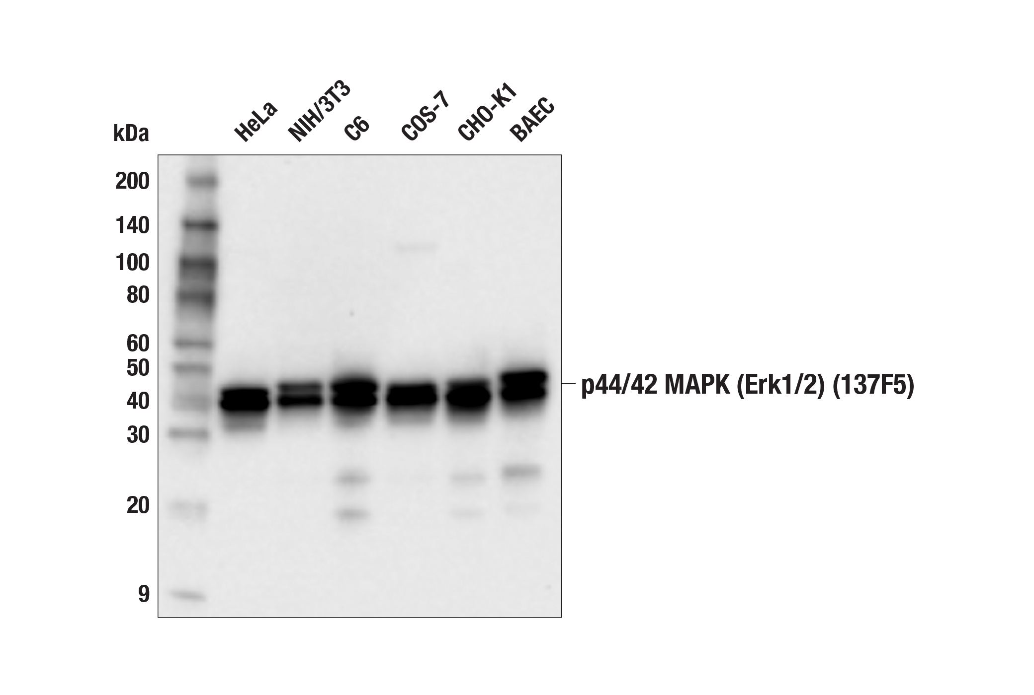
Western blot analysis of extracts from HeLa, NIH/3T3 and C6 cells, using p44/42 MAPK (Erk1/2) (137F5) Rabbit mAb.
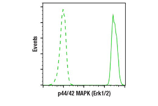
Flow cytometric analysis of Jurkat cells, U0126-treated (blue) or PMA-treated (green), using p44/42 MAPK (Erk1/2) (137F5) Rabbit mAb compared to a nonspecific negative control antibody (red).
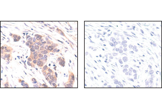
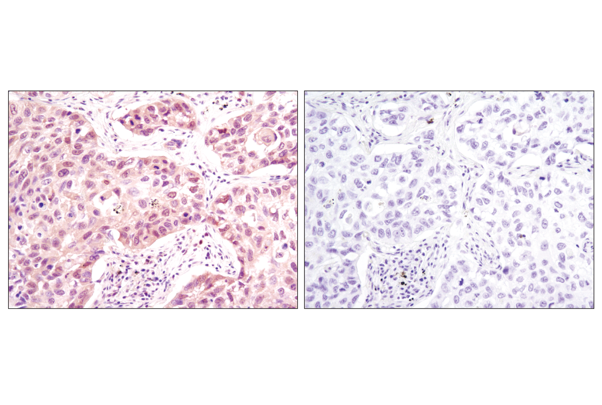
Immunohistochemical analysis of paraffin-embedded human breast carcinoma, using p44/42 MAPK (Erk1/2) (137F5) Rabbit mAb in the presence of control peptide (left) or #1240 p44/42 MAPK (Erk1/2) Blocking Peptide (#4695 Specific) (right).
Immunohistochemical analysis of paraffin-embedded human breast carcinoma using p38 MAPK (D13E1) XP® Rabbit mAb.
Western blot analysis of extracts from HeLa, NIH/3T3 and C6 cells, using p44/42 MAP Kinase (137F5) Rabbit mAb #4695.
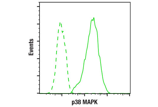
Flow cytometric analysis of HeLa cells using p38 MAPK (D13E1) XP® Rabbit mAb (blue) compared to concentration-matched Rabbit (DA1E) mAb IgG XP® Isotype Control #3900 (red).
Western blot analysis of extracts from various cell lines using p38 MAPK (D13E1) XP® Rabbit mAb #8690.
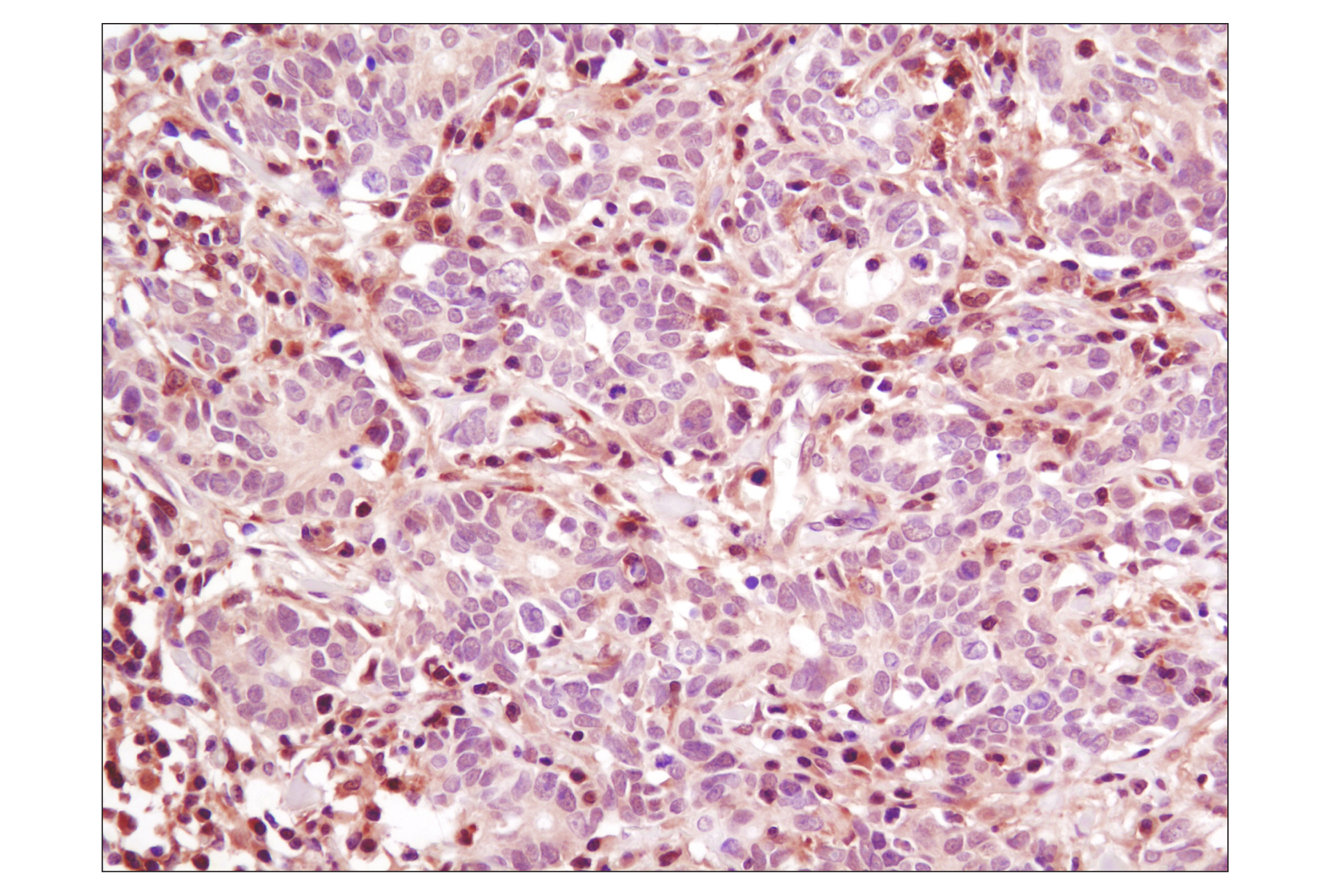
Immunohistochemical analysis of paraffin-embedded human colon carcinoma using p38 MAPK (D13E1) XP® Rabbit mAb.
After the primary antibody is bound to the target protein, a complex with HRP-linked secondary antibody is formed. The LumiGLO* is added and emits light during enzyme catalyzed decomposition.
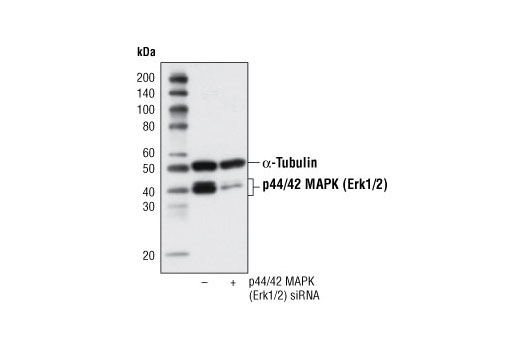
Western blot analysis of extracts from Hek 293 cells, transfected with 100 nM SignalSilence® Control siRNA (Fluorescein Conjugate) #6201 (-) or SignalSilence® p44/42 MAPK (Erk1/2) siRNA (+), using p44/42 MAPK (Erk1/2) (137F5) Rabbit mAb #4695 and α-Tubulin (11H10) Rabbit mAb #2125. The p44/42 MAPK (Erk1/2) (137F5) Rabbit mAb confirms silencing of p44/42 expression and α-Tubulin (11H10) Rabbit mAb is used to control for loading and specificity of p44/42 MAPK (Erk1/2) siRNA.
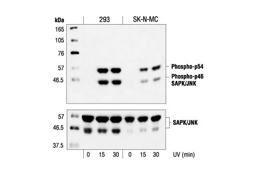
Western blot analysis of extracts from 293 and SK-N-MC cells, untreated or UV-treated (40 J/m2), using Phospho-SAPK/JNK Antibody #9251 (upper) or SAPK/JNK Antibody (lower).
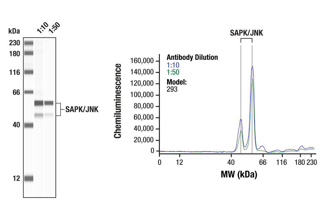
Western blot analysis of extracts from HeLa, NIH/3T3, PC12 and COS cells, using SAPK/JNK (56G8) Rabbit mAb.
Western blot analysis of extracts from HeLa, NIH/3T3, PC12 and COS cells using SAPK/JNK (56G8) Rabbit mAb #9258.
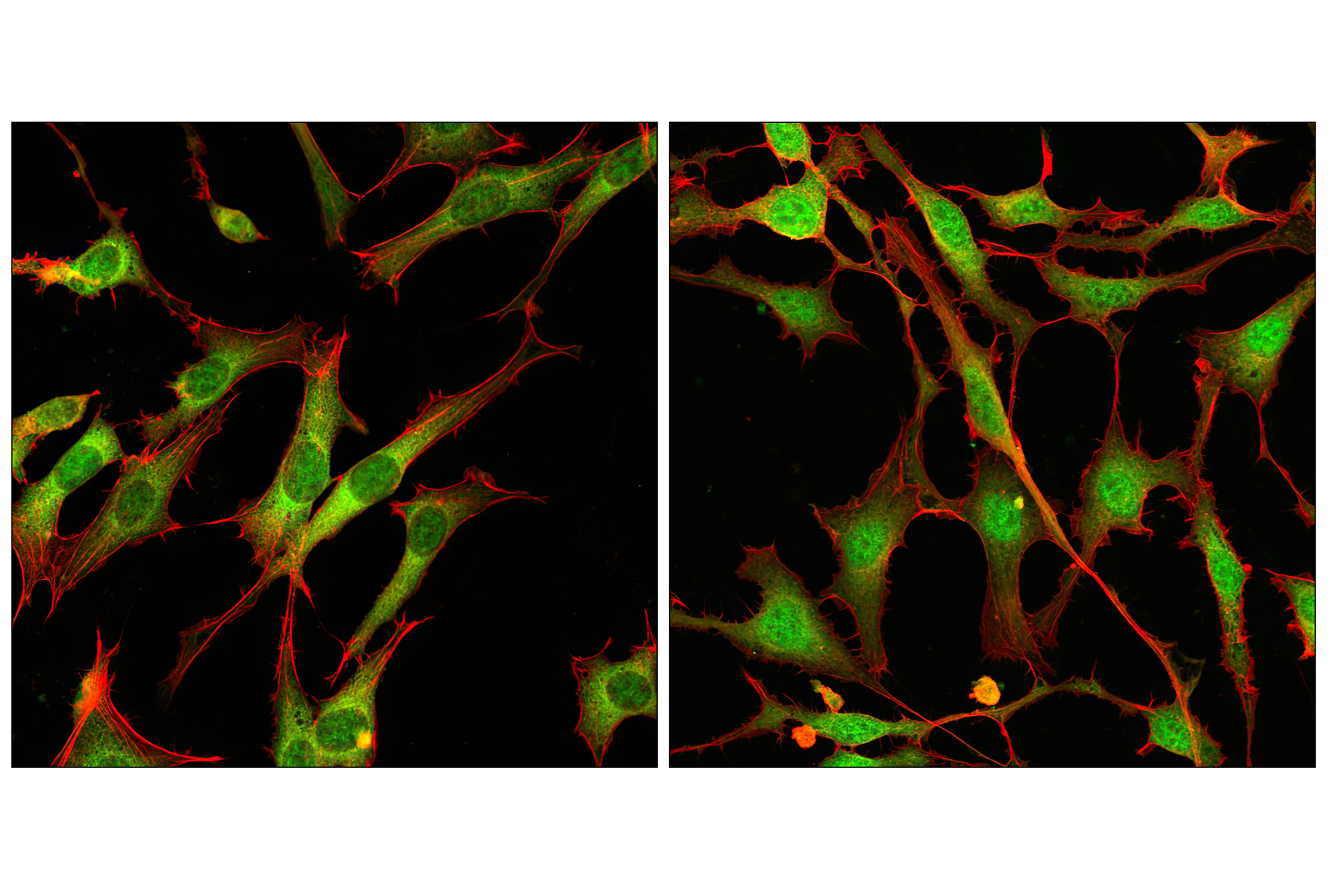
Confocal immunofluorescent analysis of NIH/3T3 cells, treated with either U0126 (MEK1/2 Inhibitor) #9903 (left) or PDGF (right), using p44/42 MAPK (Erk1/2) (137F5) Rabbit mAb (green). Actin filaments have been labeled with DY-554 phalloidin (red).
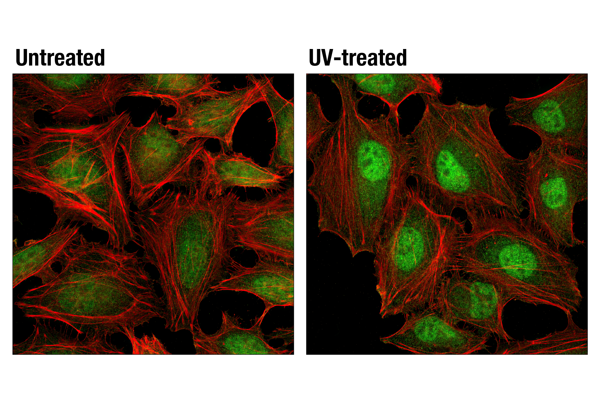
Confocal immunofluorescent analysis of HeLa cells, untreated (left) or treated with UV (100 mJ/cm2 with 30 min recovery; right), using p38 MAPK (D13E1) XP® Rabbit mAb (green). Actin filaments were labeled with DY-554 phalloidin (red).
Immunohistochemical analysis of paraffin-embedded human lung carcinoma using p38 MAPK (D13E1) XP® Rabbit mAb in the presence of control peptide (left) or antigen-specific peptide (right).

 危险品化学品经营许可证(不带存储) 许可证编号:沪(杨)应急管危经许[2022]202944(QY)
  营业执照(三证合一)