


 全部商品分类
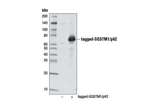


Mitophagy Antibody Sampler Kit

 下载产品说明书 下载SDS
 用小程序,查商品更便捷


 收藏
收藏
 对比
对比 咨询
咨询
The Mitophagy Antibody Sampler Kit provides an economical means of detecting proteins involved in the process of mitophagy. The kit includes enough primary antibody to perform two western blot experiments with each primary antibody.
参考图片
Western blot analysis of extracts from 293T cells, mock transfected (-) or transfected with a tagged human SQSTM1/p62 construct (+), using SQSTM1/p62 (D5E2) Rabbit mAb.
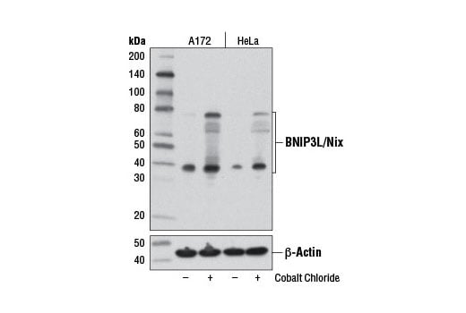
Western blot analysis of A172 and HeLa cells, untreated (-) or cobalt chloride-treated (100 μM, overnight; +), using BNIP3L/Nix (D4R4B) Rabbit mAb (upper) or β-Actin (D6A8) Rabbit mAb #8457 (lower).
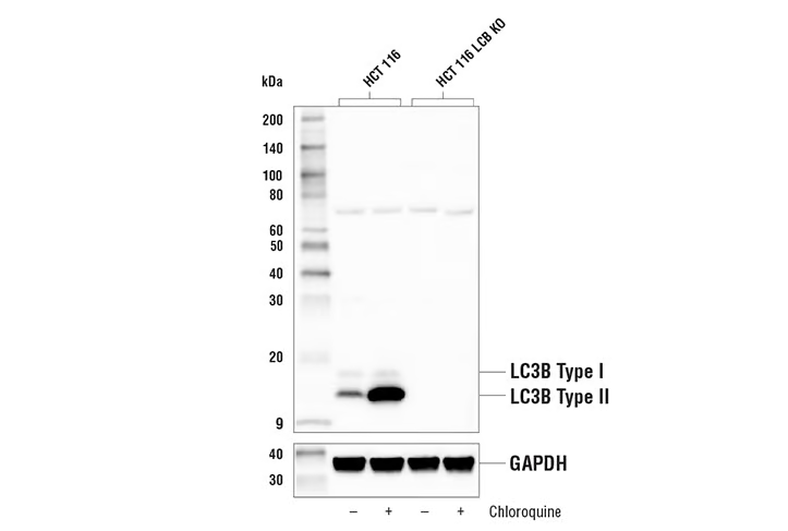
Western blot analysis of extracts from HCT 116 and HCT 116 LC3B knockout cells, untreated (-) or treated with Chloroquine #14774 (50 μM, 18 hr) using LC3B (D11) XP® Rabbit mAb #3868 (upper) or GAPDH (D16H11) XP® Rabbit mAb #5174 (lower). The absence of signal in the HCT 116 knockout cells confirms the specificity of the antibody for LC3B.
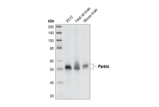
Western blot analysis of extracts from PC12 cells, fetal rat brain and mouse brain, using Parkin (Prk8) Mouse mAb.
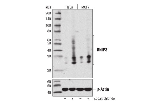
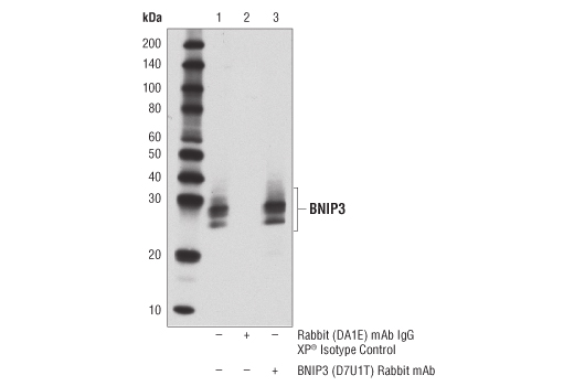
Western blot analysis of extracts from HeLa or MCF7 cells, untreated (-) or cobalt chloride-treated (100 μM, overnight; +), using BNIP3 (D7U1T) Rabbit mAb (upper) or β-Actin (D6A8) Rabbit mAb #8457 (lower).
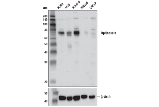
Western blot analysis of extracts from various cell lines using Optineurin (D2L8S) Rabbit mAb (upper) or β-Actin (D6A8) Rabbit mAb #8457 (lower).
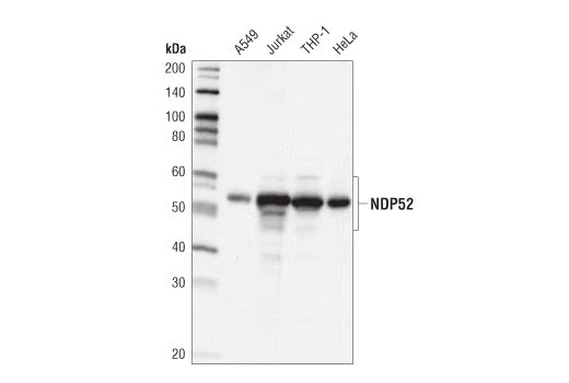
Western blot analysis of extracts from various cell lines using NDP52 (D1E4A) Rabbit mAb.
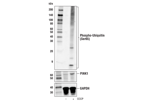
Western blot analysis of extracts from PC-3 cells, untreated (-) or treated with carbonyl cyanide 3-chlorophenylhydrazone (CCCP; 30 μM, 6 hr; +), using Phospho-Ubiquitin (Ser65) (E2J6T) Rabbit mAb (upper), PINK1 (D8G3) Rabbit mAb #6946 (middle), or GAPDH (D16H11) XP® Rabbit mAb #5174 (lower).
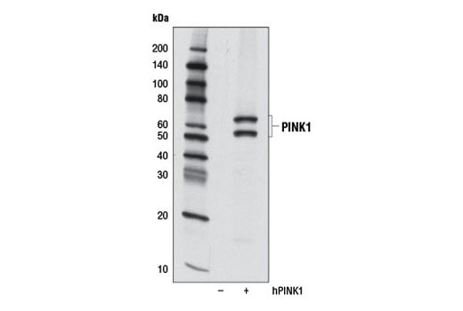
Western blot analysis of extracts from 293T cells, mock transfected (-) or transfected with a cDNA construct expressing full-length human PINK1 (hPINK1, +) using PINK1 (D8G3) Rabbit mAb.
After the primary antibody is bound to the target protein, a complex with HRP-linked secondary antibody is formed. The LumiGLO® is added and emits light during enzyme catalyzed decomposition.
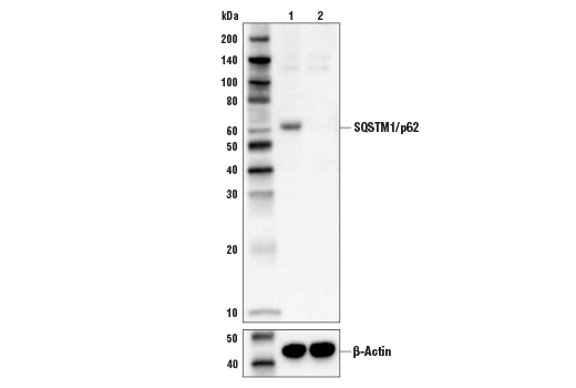
Western blot analysis of extracts from HeLa cells (lane 1) or SQSTM1 knock-out cells (lane 2) using SQSTM1/p62 (D5E2) Rabbit mAb #8025 (upper), and β-actin (D6A8) Rabbit mAb #8457 (lower). The absence of signal in the SQSTM1 knock-out HeLa cells confirms specificity of the antibody for SQSTM1.
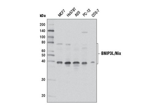
Western blot analysis of extracts from various cell lines using BNIP3L/Nix (D4R4B) Rabbit mAb.
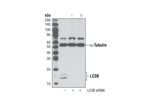
Western blot analysis of extracts from HeLa cells, transfected with 100 nM SignalSilence® Control siRNA (Unconjugated) #6568 (-), SignalSilence® LC3B siRNA I #6212 (+) or SignalSilence® LC3B siRNA II #6213 (+), using LC3B (D11) XP® Rabbit mAb #3868 and α-Tubulin (11H10) Rabbit mAb #2125. The LC3B (D11) XP® Rabbit mAb confirms silencing of LC3B expression, while the α-Tubulin (11H10) Rabbit mAb is used to control for loading and specificity of LC3B siRNA.
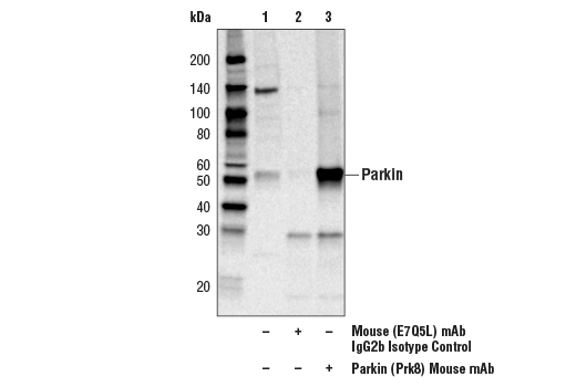
Immunoprecipitation of Parkin protein from PC12 extracts. Lane 1 is 10% input, lane 2 is Mouse (E7Q5L) mAb IgG2b Isotype Control #53484, and lane 3 is Parkin (Prk8) Mouse mAb. Western blot analysis was performed using Parkin Antibody #2132. Anti-rabbit IgG, HRP-linked Antibody #7074 was used as a secondary antibody.
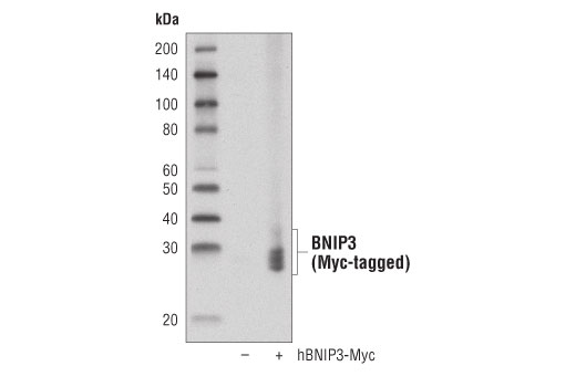
Western blot analysis of extracts from 293T cells, mock transfected (-) or transfected with a construct expressing Myc-tagged full-length human BNIP3 protein (hBNIP3-Myc; +), using BNIP3 (D7U1T) Rabbit mAb.
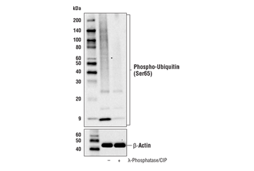
Western blot analysis of extracts from PC-3 cells treated with carbonyl cyanide 3-chlorophenylhydrazone (CCCP; 30 μM, 6 hr ). Lysates were treated with (+) or without (-) Lambda Phosphatase (λ-Phosphatase) and Calf Intestinal Phosphatase (CIP). Western blot was performed using Phospho-Ubiquitin (Ser65) (E2J6T) Rabbit mAb (upper) or β-Actin (D6A8) Rabbit mAb #8457 (lower).
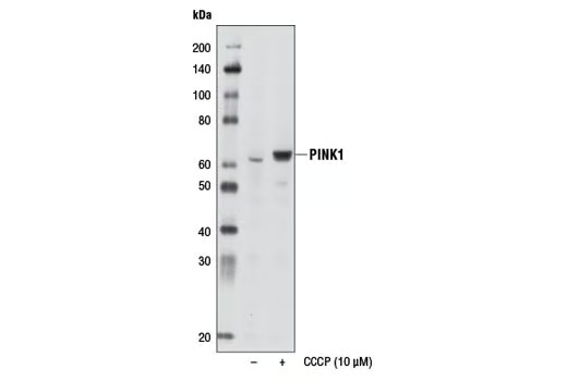
Western blot analysis of extracts from HeLa cells, untreated (-) or treated with CCCP (10 μM, 24 hr; +), using PINK1 (D8G3) Rabbit mAb.
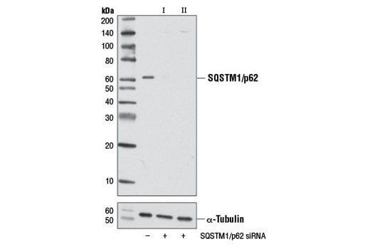
Western blot analysis of extracts from HeLa cells, transfected with 100 nM SignalSilence® Control siRNA (Unconjugated) #6568 (-), SignalSilence® SQSTM1/p62 siRNA I #6394 (+) or SignalSilence® SQSTM1/p62 siRNA II #6399 (+), using SQSTM1/p62 (D5E2) Rabbit mAb (upper) or α-Tubulin (11H10) Rabbit mAb #2125 (lower). The SQSTM1/p62 (D5E2) Rabbit mAb confirms silencing of SQSTM1/p62 expression, while the α-Tubulin (11H10) Rabbit mAb is used as a loading control.
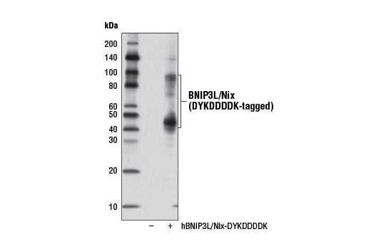
Western blot analysis of extracts from 293T cells, mock transfected (-) or transfected with a construct expressing DYKDDDDK-tagged full-length human BNIP3L/Nix (hBNIP3L/Nix-DYKDDDDK; +), using BNIP3L/Nix (D4R4B) Rabbit mAb. The BNIP3L/Nix construct was kindly provided by Dr. Reuben Shaw, Salk Institute for Biological Studies, La Jolla, CA.
Immunoprecipitation of BNIP3 from MCF7 cells treated with cobalt chloride (100 μM; overnight). Lane 1 represents 10% input, lane 2 is Rabbit (DA1E) mAb IgG XP® Isotype Control #3900, and lane 3 is BNIP3 (D7U1T) Rabbit mAb. Western blot analysis was performed using BNIP3 (D7U1T) Rabbit mAb. A conformation-specific secondary antibody was used to avoid reactivity with IgG.
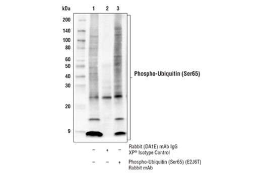
Immunoprecipitation of phospho-ubiquitin (Ser65) from extracts of PC-3 cells treated with carbonyl cyanide 3-chlorophenylhydrazone (CCCP; 30 μM, 6 hr). Lane 1 is 10% input, lane 2 is precipitated with Rabbit (DA1E) mAb IgG XP® Isotype Control #3900, and lane 3 is Phospho-Ubiquitin (Ser65) (E2J6T) Rabbit mAb. Western blot was performed using Phospho-Ubiquitin (Ser65) (E2J6T) Rabbit mAb. Mouse Anti-Rabbit IgG (Conformation Specific) (L27A9) mAb (HRP Conjugate) #5127 was used as a secondary antibody.
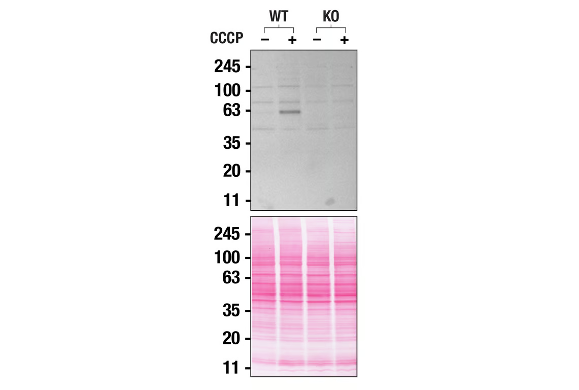
Western blot analysis of HAP1 extracts from WT or PINK1 KO untreated (-) or treated with CCCP (+) using PINK1 (D8G3) Rabbit mAb. Membranes stained with Ponceau S for total protein normalization (lower). These data were provided by YCharOS Inc., an open science company with the mission of characterizing commercially available antibodies, as a companion to validation data generated by CST scientists.
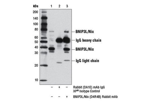
Immunoprecipitation of BNIP3L/Nix from A172 cells, treated with cobalt chloride (100 μM, overnight), using Rabbit (DA1E) mAb IgG XP® Isotype Control #3900 (lane 2) or BNIP3L/Nix (D4R4B) Rabbit mAb (lane 3). Lane 1 is 10% input. Western blot analysis was performed using BNIP3L/Nix (D4R4B) Rabbit mAb.
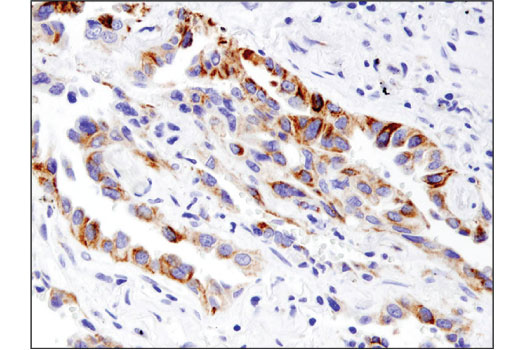
Immunohistochemical analysis of paraffin-embedded human non-small cell lung carcinoma using BNIP3 (D7U1T) Rabbit mAb.
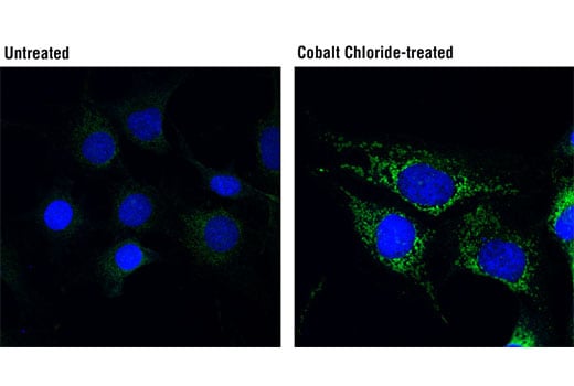
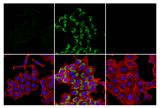
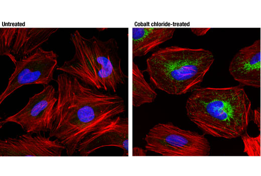
Confocal immunofluorescent analysis of A172 cells, untreated (left) or cobalt chloride-treated (100 μM, overnight; right), using BNIP3L/Nix (D4R4B) Rabbit mAb (green). Blue pseudocolor = DRAQ5® #4084 (fluorescent DNA dye).
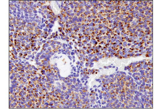
Immunohistochemical analysis of paraffin-embedded human ovarian carcinoma using BNIP3 (D7U1T) Rabbit mAb.
Confocal immunofluorescent analysis of HCT 116 cells either untreated (left) or treated with Chloroquine #14774 (50 µM, overnight) (center) or LC3B HCT 116 knockout cells treated with Chloroquine #14774 (50 µM, overnight) (right) using LC3B (D11) XP® Rabbit mAb (green). Actin filaments were labeled with β-Actin (8H10D10) Mouse mAb (red) and nuclei were labeled with DAPI #4083 (blue).
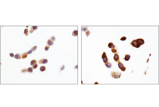
Immunohistochemical analysis of paraffin-embedded MCF7 cell pellets, untreated (left) or cobalt chloride-treated (right), using BNIP3 (D7U1T) Rabbit mAb.
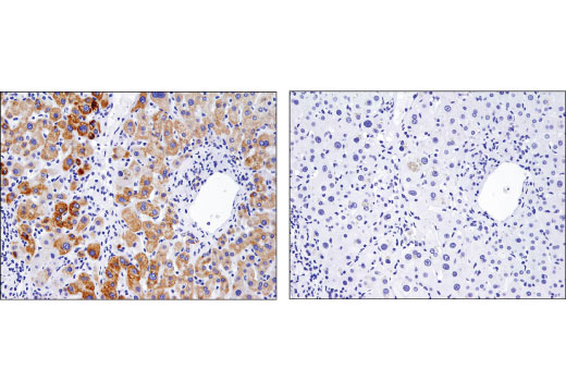
Immunohistochemical analysis of paraffin-embedded normal human liver using BNIP3 (D7U1T) Rabbit mAb in the presence of control peptide (left) and antigen-specific peptide (right).
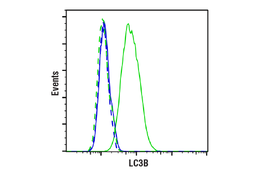
Flow cytometric analysis of HCT-116 cells, wild-type (green, high expression) or LC3B knockdown (blue, negative expression), using LC3B (D11) XP® Rabbit mAb #3868 (solid lines) or a concentration-matched Rabbit (DA1E) mAb IgG XP® Isotype Control #3900 (dashed lines). Anti-rabbit IgG (H+L), F(ab')2 Fragment (Alexa Fluor® 488 Conjugate) #4412 was used as a secondary antibody.
Confocal immunofluorescent analysis of HeLa cells, untreated (left) or treated with cobalt chloride (100 μM, 24 hr; right), using BNIP3 (D7U1T) Rabbit mAb (green). Actin filaments were labeled with DyLight™ 554 Phalliodin #13054 (red). Blue pseudocolor = DRAQ5® #4084 (fluorescent DNA dye).
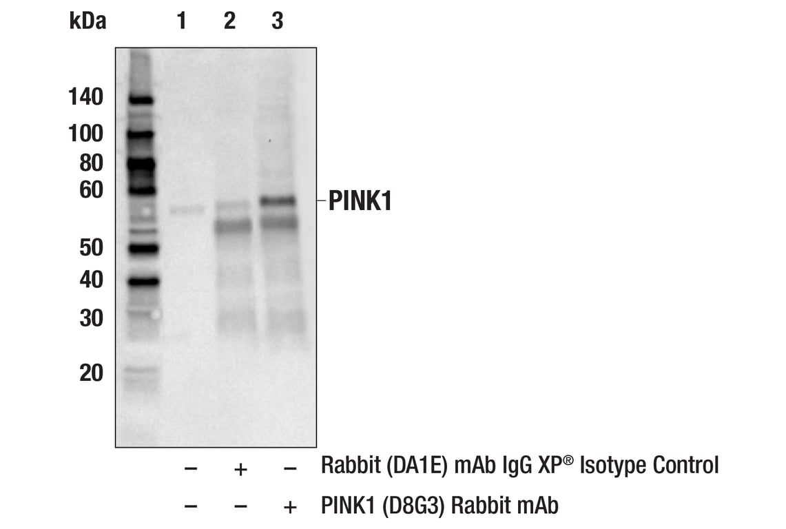
Immunoprecipitation of PINK1 protein from PC-3+CCCP extracts. Lane 1 is 10% input, lane 2 is Rabbit (DA1E) mAb IgG XP® Isotype Control, #3900 and lane 3 is PINK1 (D8G3) Rabbit mAb. Western blot analysis was performed using PINK (D8G3) Rabbit mAb, #6946. Anti-rabbit IgG, HRP-linked Antibody, #7074 was used as the secondary antibody.
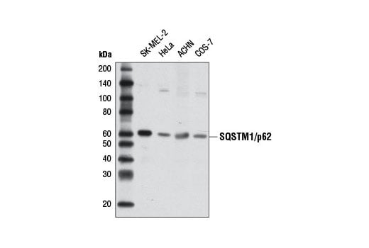
Western blot analysis of extracts from various cell lines using SQSTM1/p62 (D5E2) Rabbit mAb.
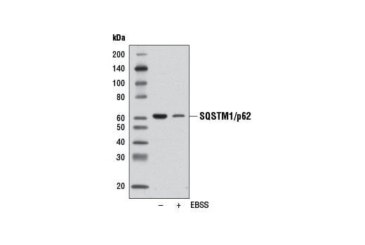
Western blot analysis of extracts from SK-MEL-2 cells, untreated (-) or starved overnight in Earle's Balanced Salt Solution (EBSS) (+), using SQSTM1/p62 (D5E2) Rabbit mAb.