


 全部商品分类
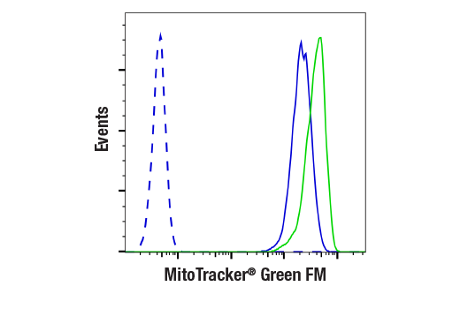


MitoTracker ® Green FM

 下载产品说明书 下载COA 下载SDS
 用小程序,查商品更便捷


 收藏
收藏
 对比
对比 咨询
咨询MitoTracker® Green FM is recommended for live cell imaging only; fixation with aldehydes or alcohols will inhibit staining. Excitation: 490 nm, Emission: 516 nm, Molecular Weight: 671.88 g/mol

Product Usage Information
Each vial of MitoTracker® Green FM contains 50 µg lyophilized solid. To make a 1 mM stock solution, reconstitute the solid in 74.4 µl high quality DMSO. While the optimal staining concentration and incubation times can vary by application, typical results are obtained by diluting the stock solution directly into normal growth media at a concentration between 100-400 nM and incubating between 15-30 min at 37ºC. After incubation, the cells must be imaged live; DO NOT FIX!To reduce background fluorescence of the media, phenol red free media can be substituted for normal media prior to imaging.
Store lyophilized solid at -20°C dessicated and protected from light. In lyophilized form, this reagent is stable for 12 months. Once reconstituted in DMSO, the solution should be stored at -20°C, protected from light, and must be used within 2 weeks. Avoid freeze-thaw cycles.
参考图片
Flow cytometric analysis of live Jurkat cells, untreated (blue) or treated with CCCP (80 μM, 15 min; green) labeled with MitoTracker® Green FM (solid lines), compared to unlabeled Jurkat cells (dashed line).
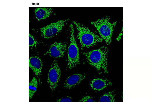
Confocal analysis of HeLa cells using MitoTracker® Green FM (green) and DRAQ5® #4084 (blue psuedocolor).