


 全部商品分类
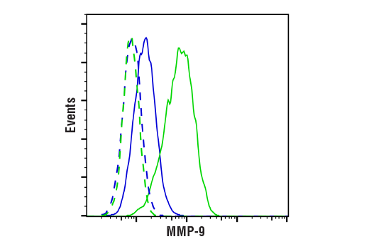


MMP-9 (D6O3H) XP® Rabbit mAb

 下载产品说明书 下载COA 下载SDS
 用小程序,查商品更便捷


 收藏
收藏
 对比
对比 咨询
咨询
Monoclonal antibody is produced by immunizing animals with a synthetic peptide corresponding to residues surrounding Phe542 of human MMP-9 protein.
Product Usage Information
| Application | Dilution |
|---|---|
| Western Blotting | 1:1000 |
| Immunohistochemistry (Paraffin) | 1:150 - 1:600 |
| Flow Cytometry (Fixed/Permeabilized) | 1:200 - 1:800 |
Specificity/Sensitivity
Species Reactivity:
Human
Supplied in 10 mM sodium HEPES (pH 7.5), 150 mM NaCl, 100 µg/ml BSA, 50% glycerol and less than 0.02% sodium azide. Store at –20°C. Do not aliquot the antibody.
For a carrier-free (BSA and azide free) version of this product see product #15749.
参考图片
Flow cytometric analysis of U-2 OS cells, untreated (blue) or treated with TPA (12-O-Tetradecanoylphorbol-13-Acetate) #4174 (200 nM, 24 hr; green) using MMP-9 (D6O3H) XP® Rabbit mAb (solid lines) or concentration-matched Rabbit (DA1E) mAb IgG XP® Isotype Control #3900 (dashed lines). Anti-rabbit IgG (H+L), F(ab')2 Fragment (Alexa Fluor® 488 Conjugate) #4412 was used as a secondary antibody.
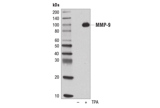
Western blot analysis of concentrated, serum-free cultured medium from U-2 OS cells, untreated (-) or treated with TPA #4174 (200 nM, 48 hr; +), using MMP-9 (D6O3H) XP® Rabbit mAb.
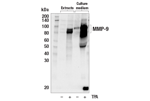
Western blot analysis of extracts and concentrated culture medium from U-2 OS cells, untreated (-) or treated with TPA #4174 (200 nM, 48 hr; +) using MMP-9 (D6O3H) XP® Rabbit mAb. MMP-9 is induced by TPA treatment as expected.
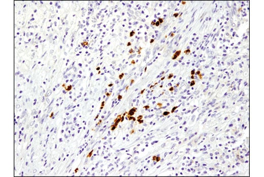
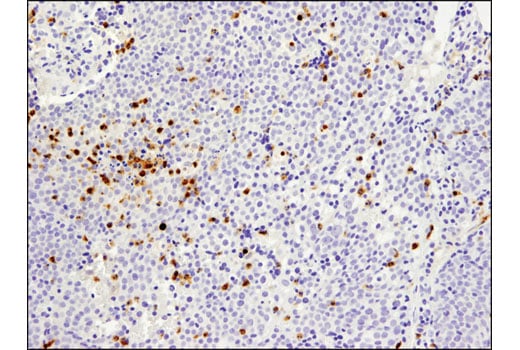
Immunohistochemical analysis of paraffin-embedded human breast carcinoma using MMP-9 (D6O3H) XP® Rabbit mAb.
Immunohistochemical analysis of paraffin-embedded human lung carcinoma using MMP-9 (D6O3H) XP® Rabbit mAb.
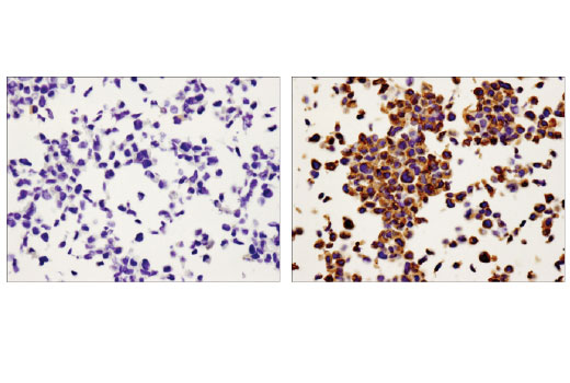
Immunohistochemical analysis of paraffin-embedded U-2 OS cell pellets, untreated (left) or treated with TPA #4174 (right), using MMP-9 (D6O3H) XP® Rabbit mAb.
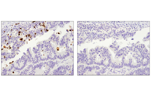
Immunohistochemical analysis of paraffin-embedded human colon carcinoma using MMP-9 (D6O3H) XP® Rabbit mAb in the presence of control peptide (left) or antigen-specific peptide (right).