


 全部商品分类


Mouse Microglia Marker IF Antibody Sampler Kit

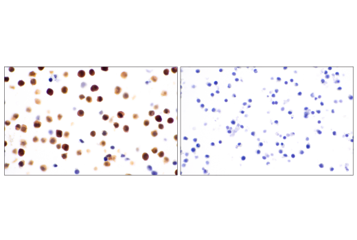
 下载产品说明书 下载SDS
 用小程序,查商品更便捷


 收藏
收藏
 对比
对比 咨询
咨询
The Mouse Microglia Marker IF Antibody Sampler Kit provides an economical means of detecting proteins identified as microglia markers by immunofluorescence and/or western blot. This kit includes enough primary antibodies to perform at least twenty IF-F tests or two western blot experiments per primary antibody.
参考图片
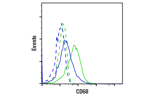
Flow cytometric analysis of RAW 264.7 cells, untreated (blue) or treated with TPA (12-O-Tetradecanoylphorbol-13-Acetate) #4174 (100 ng/mL, 24 h; green) using CD68 (E3O7V) Rabbit mAb (solid lines) or concentration-matched Rabbit (DA1E) mAb IgG XP® Isotype Control #3900 (dashed lines). Anti-rabbit IgG (H+L), F(ab')₂ Fragment (Alexa Fluor® 488 Conjugate) #4412 was used as a secondary antibody.
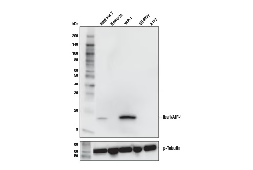
Western blot analysis of extracts from various cell lines using Iba1/AIF-1 (E4O4W) XP® Rabbit mAb (upper) and β-Tubulin (D2N5G) Rabbit mAb #15115 (lower).
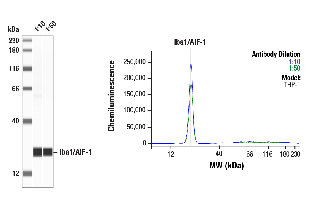
Simple Western™ analysis of lysates (1 mg/mL) from THP-1 cells using Iba1/AIF-1 (E4O4W) XP® Rabbit mAb #17198. The virtual lane view (left) shows the target band (as indicated) at 1:10 and 1:50 dilutions of primary antibody. The corresponding electropherogram view (right) plots chemiluminescence by molecular weight along the capillary at 1:10 (blue line) and 1:50 (green line) dilutions of primary antibody. This experiment was performed under reducing conditions on the Jess™ Simple Western instrument from ProteinSimple, a BioTechne brand, using the 12-230 kDa separation module.
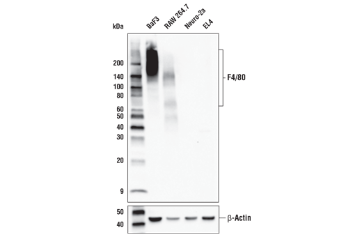
Western blot analysis of extracts from various cell lines using F4/80 (D4C8V) XP® Rabbit mAb (upper) or β-Actin (D6A8) Rabbit mAb #8457 (lower).
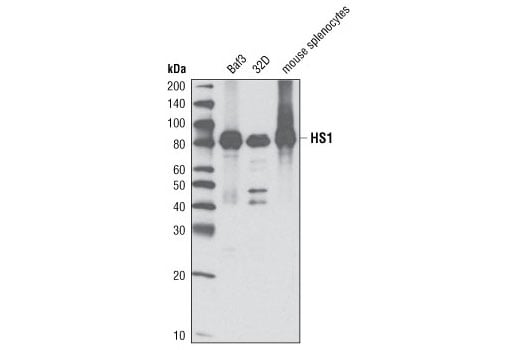
Western blot analysis of cell extracts from Baf3, 32D, and mouse spleen using HS1 (D5A9) XP® Rabbit mAb.
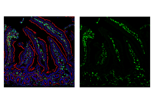
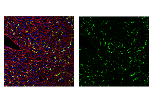
Confocal immunofluorescent analysis of mouse brain microglia (left) or peripheral macrophages of the liver (right) using CD11b/ITGAM (M1/70) Rat mAb (green). Sections were mounted in ProLong® Gold Antifade Reagent with DAPI #8961 (blue).
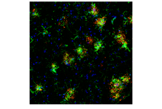
Confocal immunofluorescent analysis of brain from an amyloid mouse model of Alzheimer's Disease using CD45 (30-F11) Rat mAb (green) and β-Amyloid (D54D2) XP® Rabbit mAb #8243 (red). Sections were mounted in ProLong® Gold Antifade Reagent with DAPI #8961 (blue).
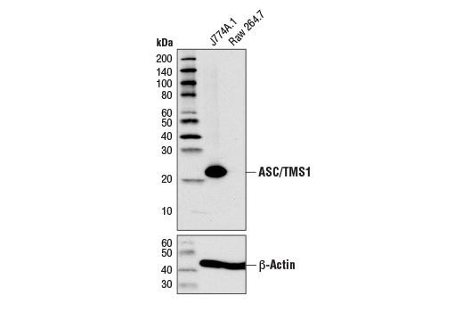
Western blot analysis of extracts from J774A.1 and Raw 264.7 cells using ASC/TMS1 (D2W8U) Rabbit mAb (upper) or β-Actin (D6A8) Rabbit mAb #8457 (lower).
After the primary antibody is bound to the target protein, a complex with HRP-linked secondary antibody is formed. The LumiGLO® is added and emits light during enzyme catalyzed decomposition.
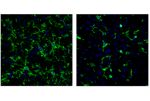
Confocal immunofluorescent analysis of wild-type mouse brain (left) and liver (right) using TMEM119 (E3E1O) Rabbit mAb (green). Sections were mounted in ProLong® Gold Antifade Reagent with DAPI #8961 (blue).
Confocal immunofluorescent analysis of the ventricular zone in P21 mouse brain using Ki-67 (D3B5) Rabbit mAb (green). Actin filaments were labeled with DyLight™ 554 phalloidin #13054 (red). Blue pseudocolor = DRAQ5® #4084 (fluorescent DNA dye).
Western blot analysis of extracts from various mouse cells using CD68 (E3O7V) Rabbit mAb (upper) or GAPDH (D16H11) XP® Rabbit mAb #5174 (lower).
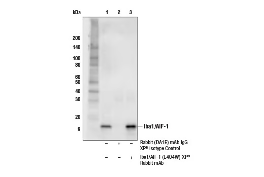
Immunoprecipitation of Iba1/AIF-1 from THP-1 cell extracts. Lane 1 is 10% input, lane 2 is Rabbit (DA1E) mAb IgG XP® Isotype Control #3900, and lane 3 is Iba1/AIF-1 (E4O4W) XP® Rabbit mAb. Western blot analysis was performed using Iba1/AIF-1 (E4O4W) XP® Rabbit mAb.
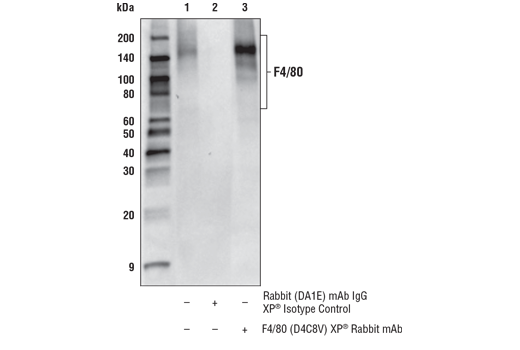
Immunoprecipitation of F4/80 from RAW 264.7 cell extracts. Lane 1 is 10% input, lane 2 is Rabbit (DA1E) mAb IgG XP® Isotype Control #3900, and lane 3 is F4/80 (D4C8V) XP® Rabbit mAb. Western blot analysis was performed using F4/80 (D4C8V) XP® Rabbit mAb as the primary antibody and Mouse Anti-Rabbit IgG (Conformation Specific) (L29A9) mAb #3678, followed by Anti-mouse IgG, HRP-linked Antibody #7076 as the secondary antibodies.
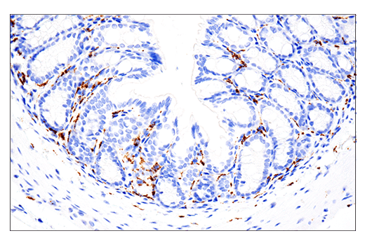
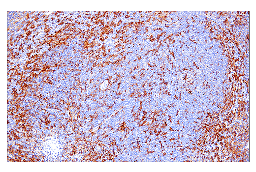
Immunohistochemical analysis of paraffin-embedded mouse spleen using HS1 (D5A9) XP® Rabbit mAb.
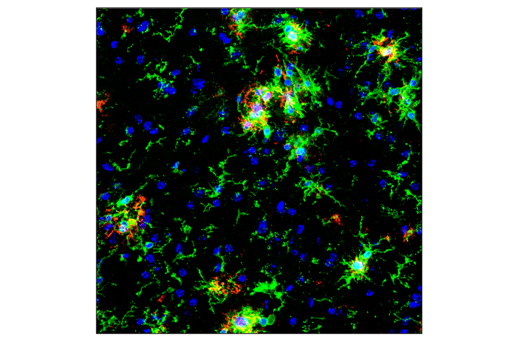
Confocal immunofluorescent analysis of brain from an amyloid mouse model of Alzheimer’s disease using CD11b/ITGAM (M1/70) Rat mAb (green) and β-Amyloid (D54D2) XP® Rabbit mAb #8243 (red). Sections were mounted in ProLong® Gold Antifade Reagent with DAPI #8961 (blue).
Confocal immunofluorescent analysis of mouse brain microglia (left) or peripheral macrophages of the liver (right) using CD45 (30-F11) Rat mAb (green). Sections were mounted in ProLong® Gold Antifade Reagent with DAPI #8961 (blue).
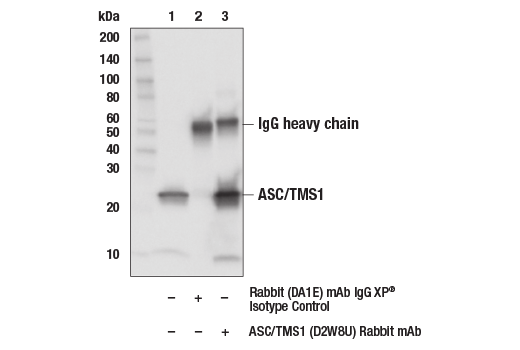
Immunoprecipitation of ASC/TMS1 from J774A.1 cell extracts. Lane 1 is 10% input, lane 2 is Rabbit (DA1E) mAb IgG XP® Isotype Control #3900, and lane 3 is ASC (D2W8U) Rabbit mAb. Western blot analysis was performed using ASC/TMS1 (D2W8U) Rabbit mAb.
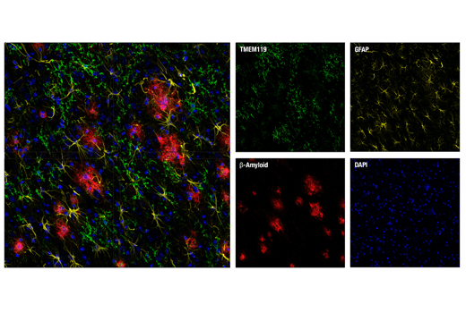
Confocal immunofluorescent analysis of brain from an amyloid mouse model of Alzheimer’s disease. Sections were labeled with TMEM119 (E3E1O) Rabbit mAb (green) and GFAP (GA5) Mouse mAb #3670 (yellow). Plaques were then stained with β-Amyloid (D54D2) XP® Rabbit mAb (Alexa Fluor® 647 Conjugate) #42284 (red) after blocking free secondary binding sites with Rabbit (DA1E) mAb IgG XP® Isotype Control #3900. Sections were mounted in ProLong® Gold Antifade Reagent with DAPI #8961 (blue).
Confocal immunofluorescent analysis of HeLa cells using Ki-67 (D3B5) Rabbit mAb (green). Actin filaments were labeled with DY-554 phalloidin (red). Blue pseudocolor = DRAQ5® #4084 (fluorescent DNA dye).
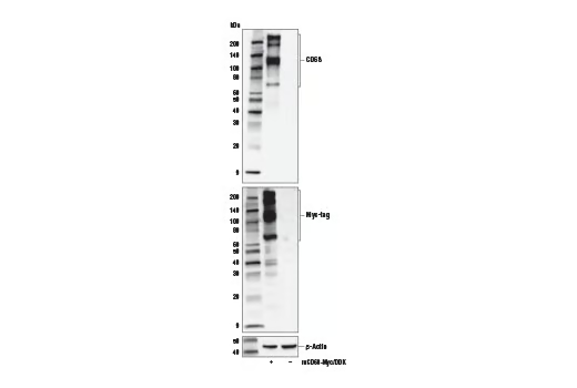
Western blot analysis of extracts from 293T cells, mock transfected (-) or transfected with a construct expressing Myc/DDK-tagged full-length mouse CD68 protein (mCD68-Myc/DDK; +), using CD68 (E3O7V) Rabbit mAb (upper), Myc-Tag (71D10) Rabbit mAb #2278 (middle), and β-Actin (D6A8) Rabbit mAb #8457 (lower).
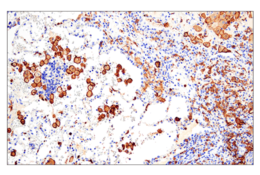
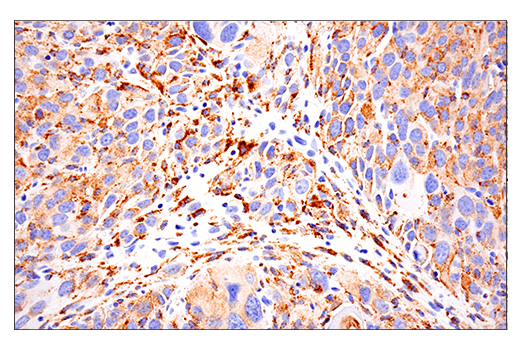
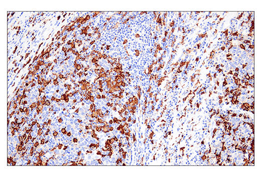
Immunohistochemical analysis of paraffin-embedded human neuroendocrine carcinoma of the lung using Iba1/AIF-1 (E4O4W) XP® Rabbit mAb performed on the Leica® BOND™ Rx.
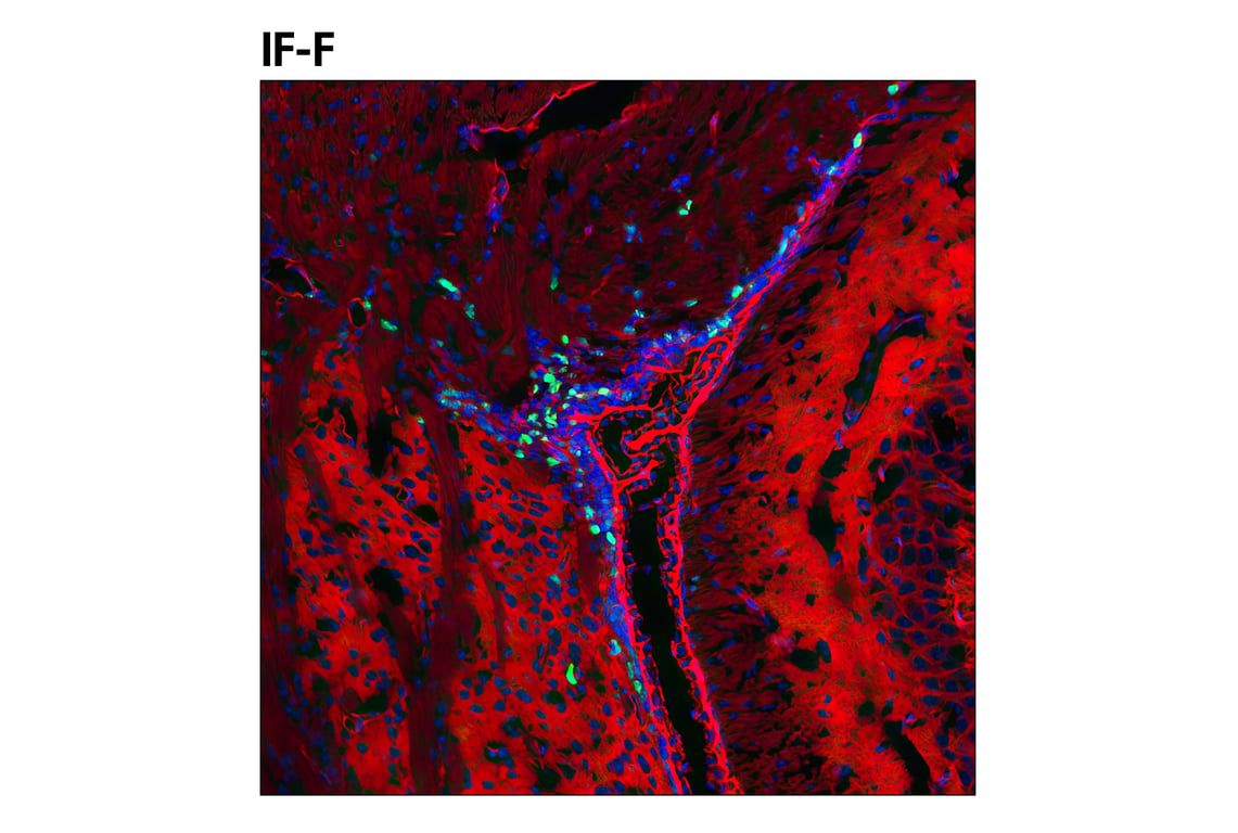
Confocal immunofluorescent analysis of Tg2576 brain, which overexpresses mutant human APP695. Sections were labeled with F4/80 (D4C8V) XP® Rabbit mAb (green), β-Amyloid (D3D2N) Mouse mAb #15126 (yellow), and GFAP (E6N9L) Mouse mAb #34001 (red). Samples were mounted in ProLong® Gold Antifade Reagent with DAPI #8961 (blue).
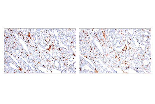
Immunohistochemical analysis of paraffin-embedded LL2 syngeneic tumor using HS1 (D5A9) XP® Rabbit mAb.
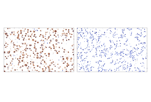
Immunohistochemical analysis of paraffin-embedded J774A.1 cell pellet (left, positive) or RAW 264.7 cell pellet (right, negative) using ASC/TMS1 (D2W8U) Rabbit mAb.
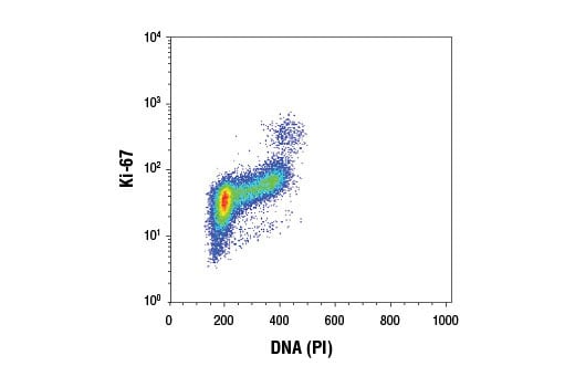
Flow cytometric analysis of Jurkat cells using Ki-67 (D3B5) Rabbit mAb and Propidium Iodide (PI)/RNase Staining Solution #4087. Anti-rabbit IgG (H+L), F(ab')2 Fragment (Alexa Fluor® 488 Conjugate) #4412 was used as a secondary antibody.
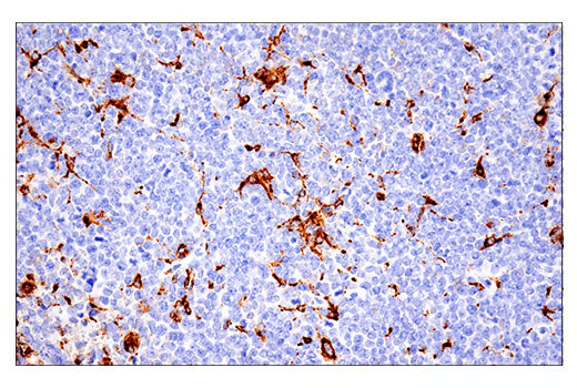
Immunohistochemical analysis of paraffin-embedded mouse spleen using CD68 (E3O7V) Rabbit mAb performed on the Leica® BOND™ Rx.
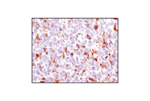
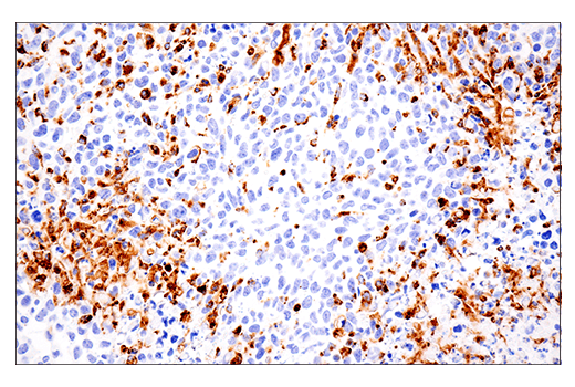
Immunohistochemical analysis of paraffin-embedded human non-Hodgkin's lymphoma using Iba1/AIF-1 (E4O4W) XP® Rabbit mAb performed on the Leica® BOND™ Rx.
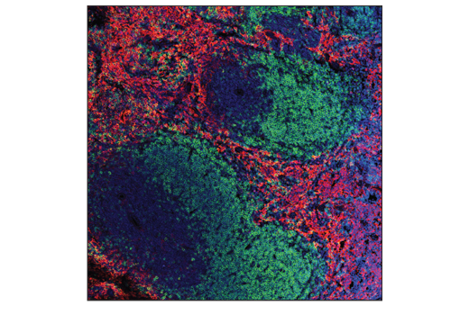
Confocal immunofluorescent analysis of normal mouse spleen using F4/80 (D4C8V) XP® Rabbit mAb (red) and B220/CD45R Alexa Fluor® 488 conjugate (green). Samples were mounted in ProLong® Gold Antifade Reagent with DAPI #8961 (blue).
Confocal immunofluorescent analysis of fixed frozen mouse cortex from wild-type (left) or an amyloid mouse model of Alzheimer's disease (right) using HS1 (D5A9) XP® Rabbit mAb (green). After blocking free secondary antibody binding sites with Rabbit (DA1E) mAb IgG XP® Isotype Control #3900, the tissue was then labeled using β-Amyloid (D54D2) XP® Rabbit mAb (Alexa Fluor® 594 Conjugate) #35363 (red) and ProLong® Gold Antifade Reagent with DAPI #8961 (blue).
Immunohistochemical analysis of paraffin-embedded mouse forestomach using ASC/TMS1 (D2W8U) Rabbit mAb.
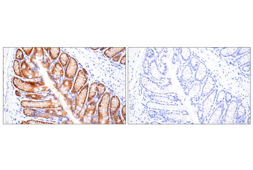
Immunohistochemical analysis of paraffin-embedded mouse colon using CD68 (E3O7V) Rabbit mAb performed on the Leica® BOND™ Rx.
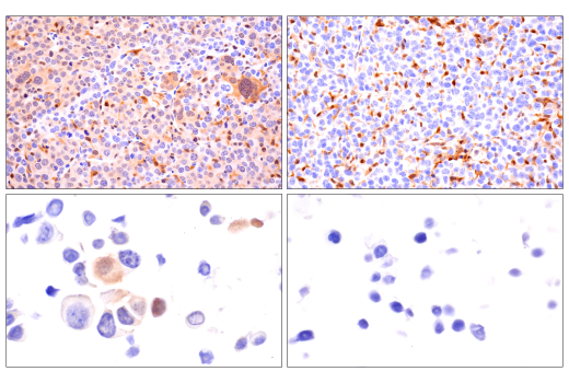
Immunohistochemical analysis of paraffin-embedded human ovarian clear cell carcinoma using Iba1/AIF-1 (E4O4W) XP® Rabbit mAb (left) or Iba1/AIF-1 (E5N4J) Mouse mAb (IHC Formulated) #58970 (right) performed on the Leica® BOND™ Rx. These two antibodies detect independent, unique epitopes on human Iba1/AIF-1 protein. The similar staining patterns obtained with both antibodies help to confirm the specificity of the staining.
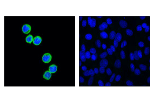
Confocal immunofluorescent analysis of RAW 264.7 cells (left, positive) or Neuro-2a cells (right, negative), using F4/80 (D4C8V) XP® Rabbit mAb (green). Samples were mounted in ProLong® Gold Antifade Reagent with DAPI #8961 (blue).
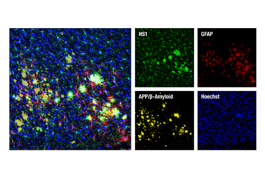
Confocal immunofluorescent analysis of mouse Tg2576 brain which overexpresses mutant human APP695. Sections were first labeled with HS1 (D5A9) XP® Rabbit mAb (green) and APP/β-Amyloid (NAB228) Mouse mAb #2450 (yellow). After blocking free secondary binding sites with Mouse (G3A1) mAb IgG1 Isotype Control #5415, sections were incubated with GFAP (GA5) Mouse mAb (Alexa Fluor® 647 Conjugate) #3657 (red). Nuclei were labeled with Hoechst 33342 #4082 (blue).
Immunohistochemical analysis of paraffin-embedded mouse brain using ASC/TMS1 (D2W8U) Rabbit mAb.
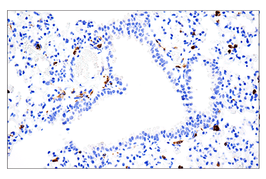
Immunohistochemical analysis of paraffin-embedded mouse lung using CD68 (E3O7V) Rabbit mAb performed on the Leica® BOND™ Rx.
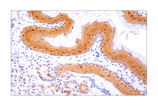
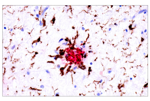
Dual immunohistochemical analysis of paraffin-embedded human Alzheimer's brain using Iba1/AIF-1 (E4O4W) XP® Rabbit mAb (brown) and APP/β-Amyloid (NAB228) Mouse mAb #2450 (red).
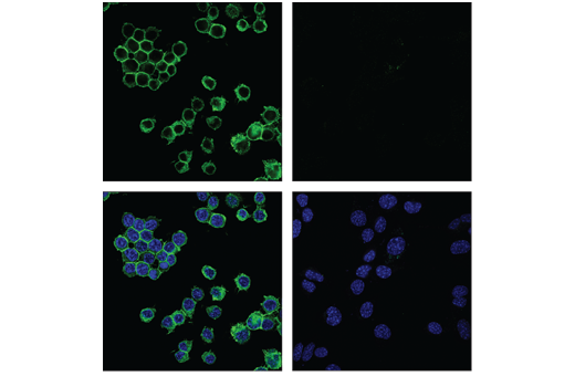
Confocal immunofluorescent analysis of 32D cells (left) and C2C12 cells (right), using HS1 (D5A9) XP® Rabbit mAb (green). Blue pseudocolor = DRAQ5® #4084 (fluorescent DNA dye).
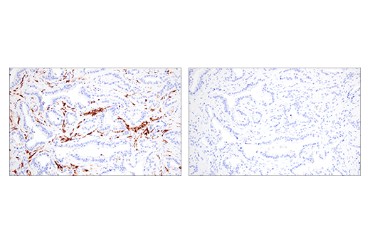
Immunohistochemical analysis of paraffin-embedded mouse colon using ASC/TMS1 (D2W8U) Rabbit mAb (left) compared to concentration-matched Rabbit (DA1E) mAb IgG XP® Isotype Control #3900 (right).
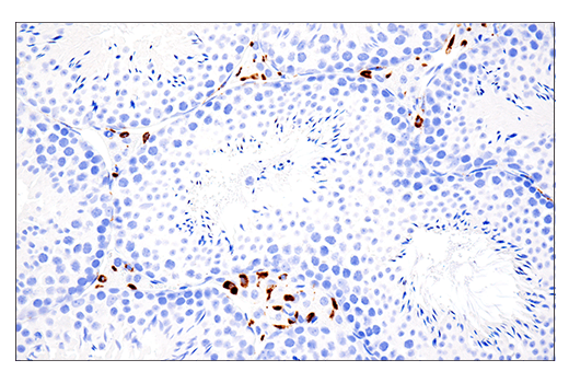
Immunohistochemical analysis of paraffin-embedded mouse testis using CD68 (E3O7V) Rabbit mAb performed on the Leica® BOND™ Rx.
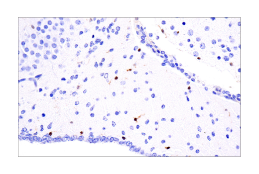
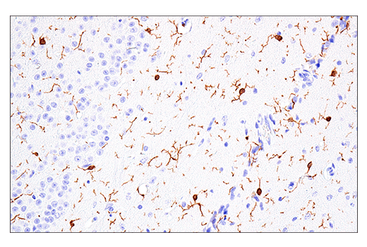
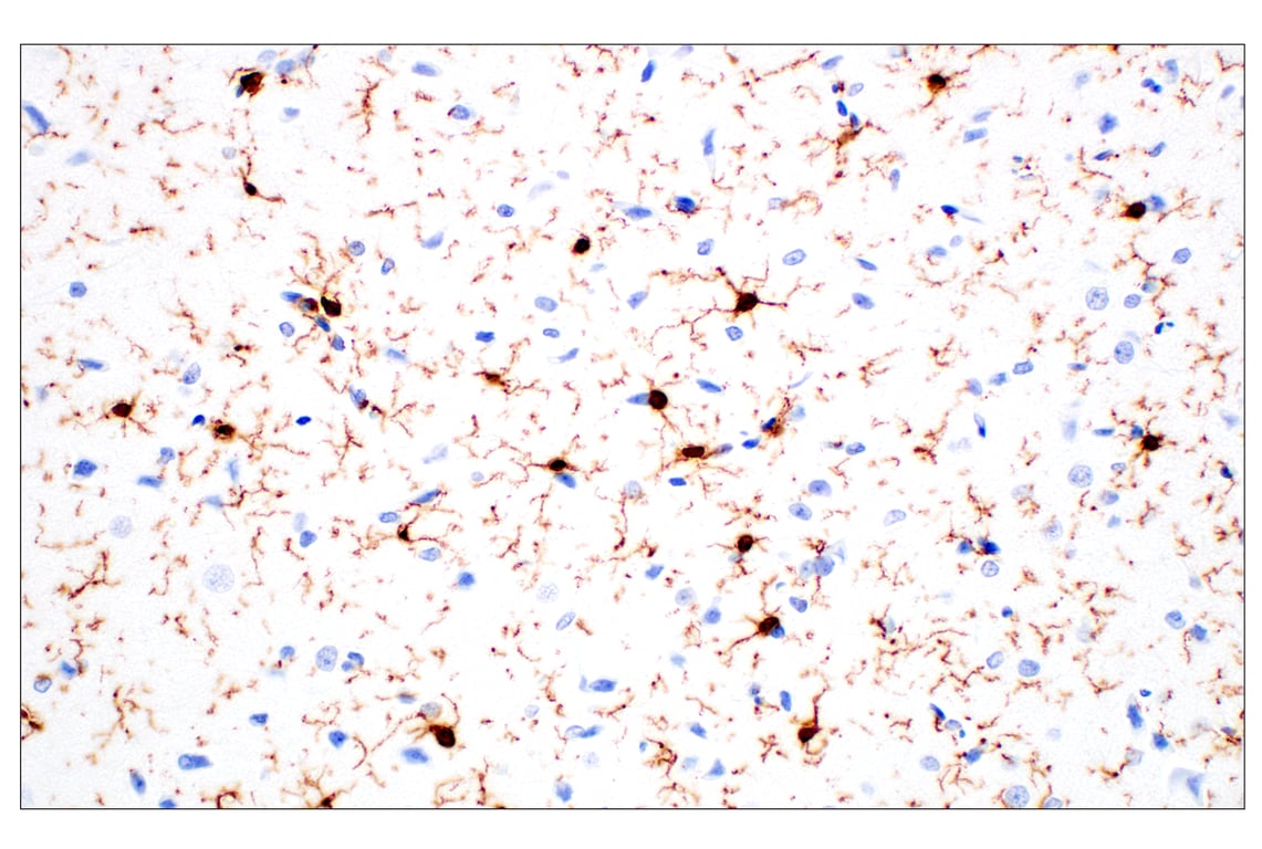
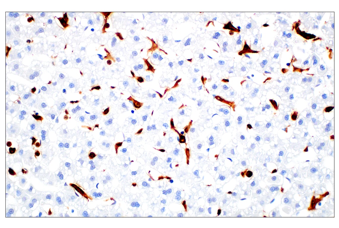
Immunohistochemical analysis of paraffin-embedded mouse brain using Iba1/AIF-1 (E4O4W) XP® Rabbit mAb.
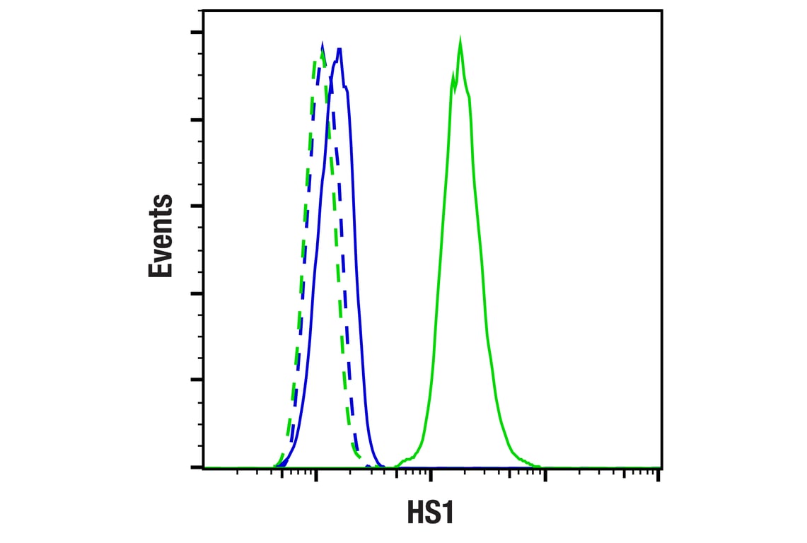
Flow cytometric analysis of NIH/3T3 cells (blue, negative) and 32D clone 3 cells (green, positive) using HS1 (D5A9) XP® Rabbit mAb (solid lines) or concentration-matched Rabbit (DA1E) mAb IgG XP® Isotype Control #3900 (dashed lines). Anti-rabbit IgG (H+L), F(ab')2 Fragment (Alexa Fluor® 488 Conjugate) #4412 was used as a secondary antibody.
Immunohistochemical analysis of paraffin-embedded mouse thymus using ASC/TMS1 (D2W8U) Rabbit mAb.
Immunohistochemical analysis of paraffin-embedded A20 syngeneic tumor using CD68 (E3O7V) Rabbit mAb.
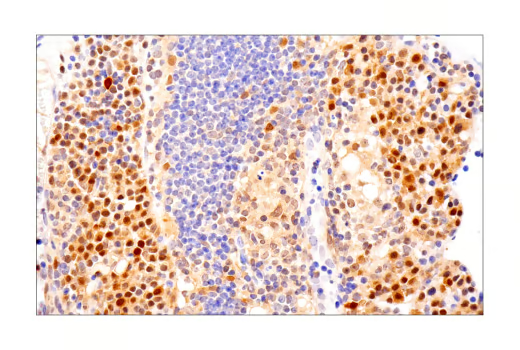
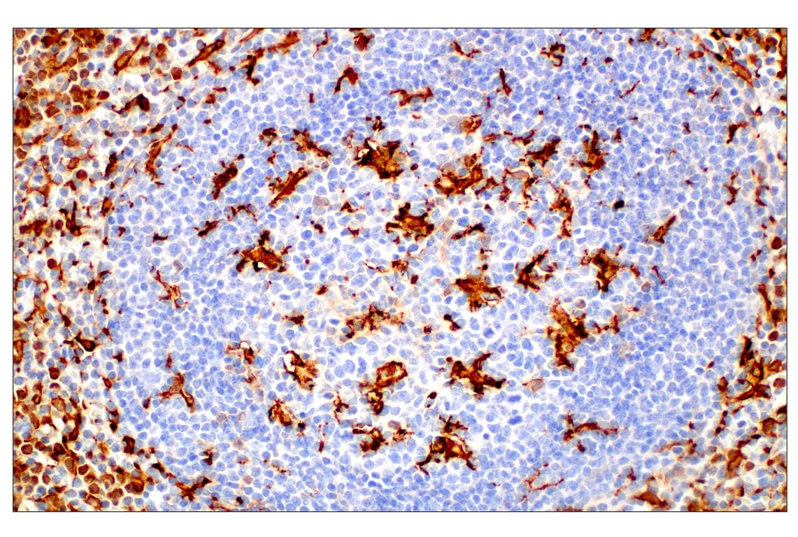
Immunohistochemical analysis of paraffin-embedded mouse spleen using Iba1/AIF-1 (E4O4W) XP® Rabbit mAb.
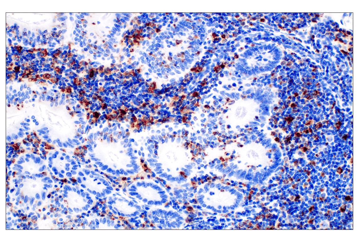
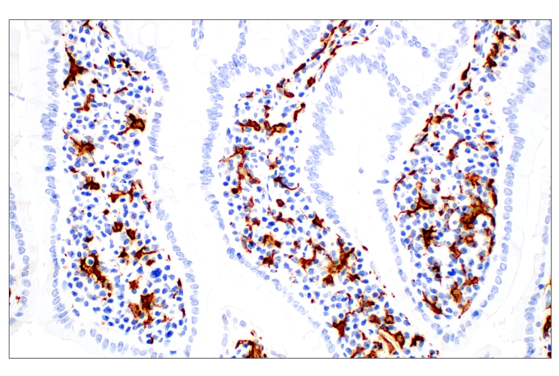
Immunohistochemical analysis of paraffin-embedded mouse small intestine using ASC/TMS1 (D2W8U) Rabbit mAb.
Immunohistochemical analysis of paraffin-embedded 4T1 syngeneic mammary tumor using CD68 (E3O7V) Rabbit mAb.
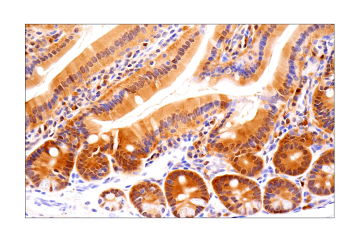
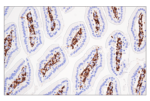
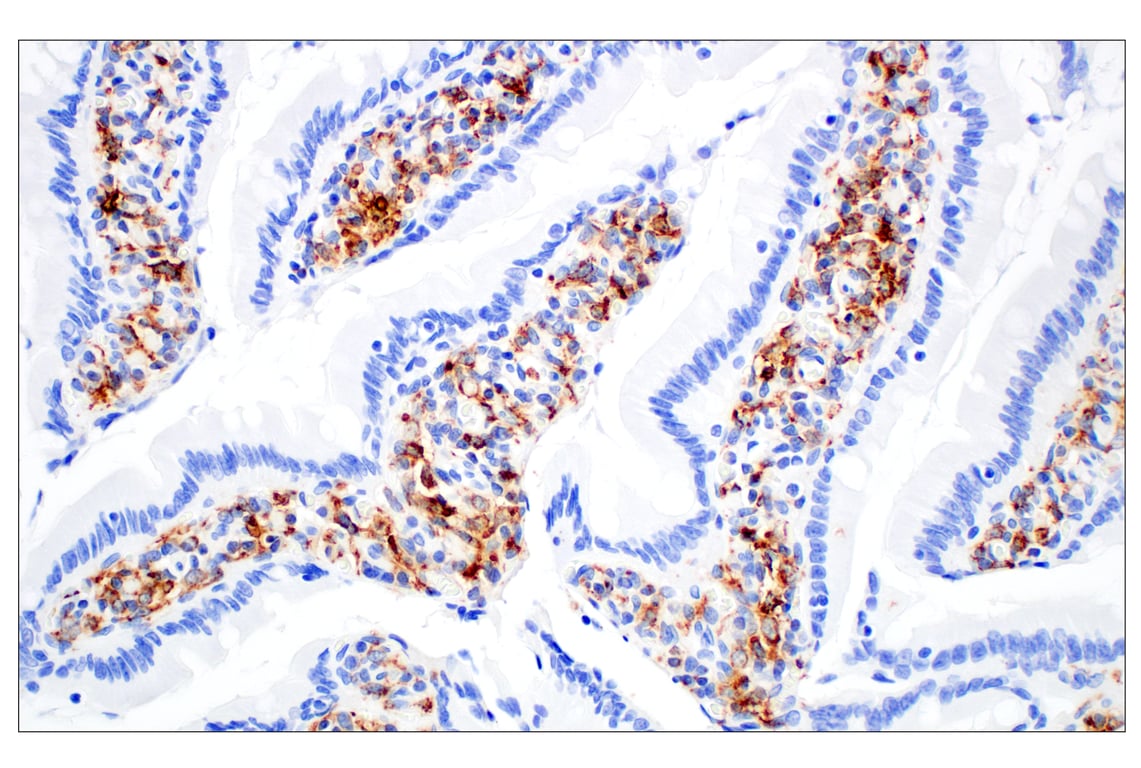
Immunohistochemical analysis of paraffin-embedded mouse small intestine using Iba1/AIF-1 (E4O4W) XP® Rabbit mAb.
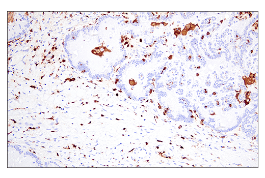
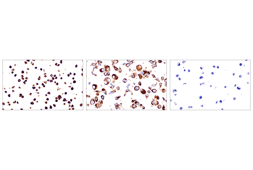
Immunohistochemical analysis of paraffin-embedded Renca syngeneic tumor (top left), 4T1 syngeneic mammary tumor (top right), Renca cell pellet (bottom left), and 4T1 cell pellet (bottom right) using ASC/TMS1 (D2W8U) Rabbit mAb. Both tumors show staining of infiltrating immune cells. Note the presence of staining in the Renca tumor cells and the lack of staining in the 4T1 tumor cells consistent with staining results on corresponding cell pellets.
Immunohistochemical analysis of paraffin-embedded Renca syngeneic tumor using CD68 (E3O7V) Rabbit mAb.
Immunohistochemical analysis of paraffin-embedded normal rat brain using Iba1/AIF-1 (E4O4W) XP® Rabbit mAb.
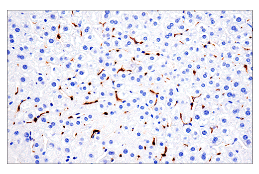
Immunohistochemical analysis of paraffin-embedded mouse liver using CD68 (E3O7V) Rabbit mAb.
Immunohistochemical analysis of paraffin-embedded normal rhesus monkey spleen using Iba1/AIF-1 (E4O4W) XP® Rabbit mAb.
Immunohistochemical anaylsis of paraffin-embedded rat small intestine using CD68 (E3O7V) Rabbit mAb.
Immunohistochemical analysis of paraffin-embedded normal rhesus monkey liver using Iba1/AIF-1 (E4O4W) XP® Rabbit
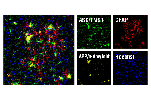
Confocal immunofluorescent analysis of mouse Tg2576 brain which overexpresses mutant human APP695. Sections were first labeled with ASC/TMS1 (D2W8U) Rabbit mAb #67824 (green) and APP/β-Amyloid (NAB228) Mouse mAb #2450 (yellow). After blocking free secondary binding sites with Mouse (G3A1) mAb IgG1 Isotype Control #5415, sections were incubated with GFAP (GA5) Mouse mAb (Alexa Fluor® 647 Conjugate) #3657 (red). Nuclei were labeled with Hoechst 33342 #4082 (blue).
Immunohistochemical anaylsis of paraffin-embedded Syrian hamster colon using CD68 (E3O7V) Rabbit mAb.
Confocal immunofluorescent analysis of human cortex (left) and mouse CA1 hippocampus (right) using Iba1/AIF-1 (E4O4W) XP® Rabbit mAb (green). In mouse tissue sections, cell nuclei were labeled with DAPI (blue). Images kindly provided by Dr. Simone Brioschi and Dr. Marco Colonna (Washington University) and used with permission.
Immunohistochemical analysis of paraffin-embedded normal Syrian hamster small intestine using Iba1/AIF-1 (E4O4W) XP® Rabbit mAb.
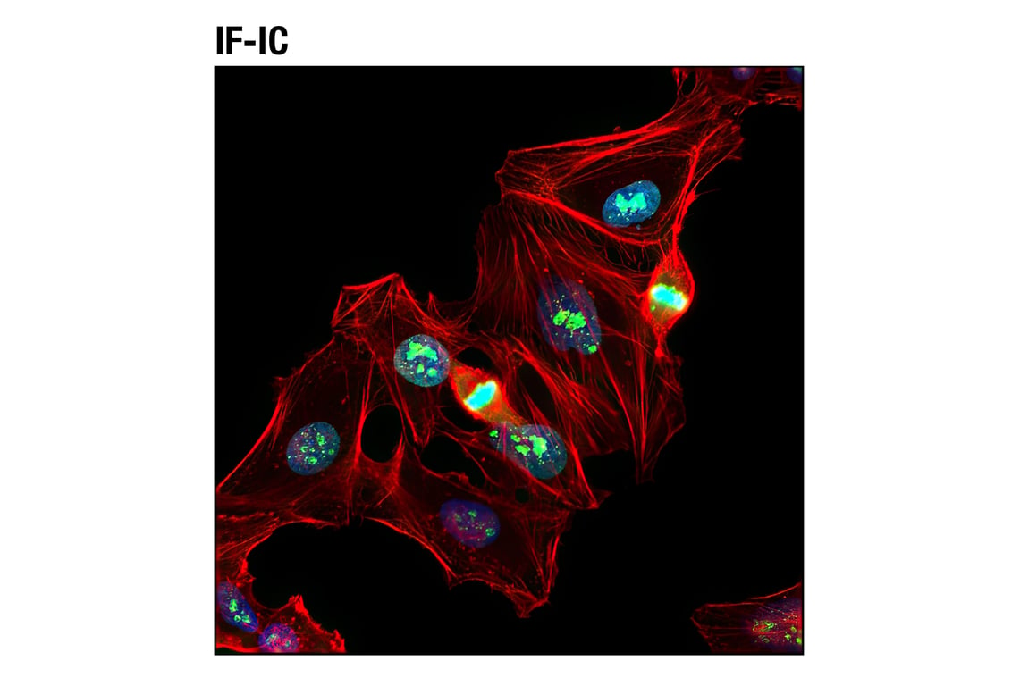
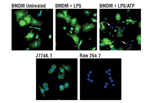
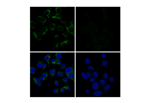
Confocal immunofluorescent analysis of mouse primary bone marrow-derived macrophages (BMDMs) either untreated (upper left) or treated with LPS (50 ng/ml, 4 hr, middle) or LPS followed by ATP (5 mM, 45 min, upper right), and J774A.1 (lower left) or Raw 264.7 (lower right) cells, using ASC/TMS1 (D2W8U) Rabbit mAb (green). Blue pseudocolor = DRAQ5® #4084 (fluorescent DNA dye). Note the translocation of ASC to inflammasomes following stimulation with LPS and ATP (white arrows).
Immunohistochemical analysis of paraffin-embedded mouse small intestine using CD68 (E3O7V) Rabbit mAb (left) compared to concentration-matched Rabbit (DA1E) mAb IgG XP® Isotype Control #3900 (right).
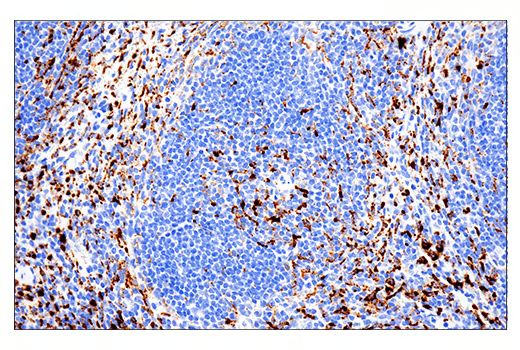
Confocal immunofluorescent analysis of mouse colon (left) and liver (right) using CD68 (E3O7V) Rabbit mAb (red). After blocking free secondary antibody binding sites with Rabbit (DA1E) mAb IgG XP® Isotype Control #3900, the tissue was then labeled using HS1 (D5A9) XP® Rabbit mAb (Rodent Specific) (Alexa Fluor® 488 Conjugate) #68206 (green). Sections were mounted in ProLong® Gold Antifade Reagent with DAPI #8961 (blue).
Immunohistochemical analysis of paraffin-embedded human ovarian serous carcinoma using Iba1/AIF-1 (E4O4W) XP® Rabbit mAb.
Confocal immunofluorescent analysis of mouse small intestine using Iba1/AIF-1 (E4O4W) XP® Rabbit mAb (green). Actin filaments were labeled with DyLight™ 554 Phalloidin #13054 (red). Sections were mounted in ProLong® Gold Antifade Reagent with DAPI #8961 (blue).
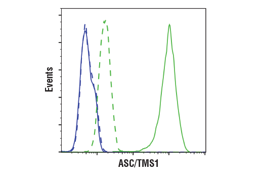
Flow cytometric analysis of Raw264.7 cells (blue) and J774A.1 cells (green) using ASC/TMS1 (D2W8U) Rabbit mAb (solid lines) or a concentration-matched Rabbit (DA1E) mAb IgG XP® Isotype Control #3900 (dashed lines). Anti-rabbit IgG (H+L), F(ab')2 Fragment (Alexa Fluor® 488 Conjugate) #4412 was used as a secondary antibody.
Immunohistochemical analysis of paraffin-embedded RAW 264.7 cell pellet (left, positive), Renca cell pellet (middle, positive), or Neuro-2a cell pellet (right, negative) using CD68 (E3O7V) Rabbit mAb.
Confocal immunofluorescent analysis of RAW 264.7 cells (left, positive) and Neuro-2a cells (right, negative) using CD68 (E3O7V) Rabbit mAb (green). Cells were mounted in ProLong® Gold Antifade Reagent with DAPI #8961 (blue).
Immunohistochemical analysis of paraffin-embedded human colon carcinoma using Iba1/AIF-1 (E4O4W) XP® Rabbit mAb.
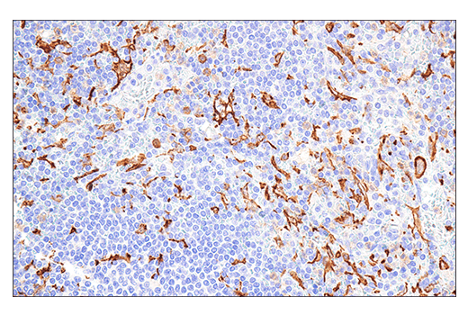
Confocal immunofluorescent analysis of mouse liver using Iba1/AIF-1 (E4O4W) XP® Rabbit mAb (green). Actin filaments were labeled with DyLight™ 554 Phalloidin #13054 (red). Sections were mounted in ProLong® Gold Antifade Reagent with DAPI #8961 (blue).
Immunohistochemical analysis of paraffin-embedded human ductal breast carcinoma using Iba1/AIF-1 (E4O4W) XP® Rabbit mAb (left) compared to concentration-matched Rabbit (DA1E) mAb IgG XP® Isotype Control #3900 (right).
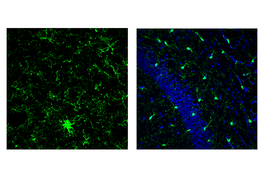
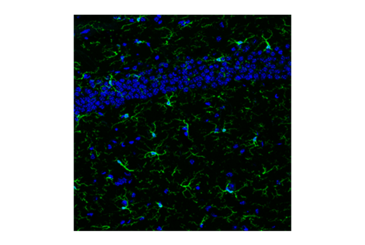
Confocal immunofluorescent analysis of microglia in mouse hippocampus using Iba1/AIF-1 (E4O4W) XP® Rabbit mAb (green). Sections were mounted in ProLong® Gold Antifade Reagent with DAPI #8961 (blue).
Immunohistochemical analysis of paraffin-embedded THP-1 cell pellet (left, positive) or SH-SY5Y cell pellet (right, negative) using Iba1/AIF-1 (E4O4W) XP® Rabbit mAb.
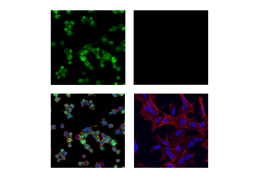
Confocal immunofluorescent analysis of THP-1 cells differentiated with TPA (12-O-Tetradecanoylphorbol-13-Acetate) #4174 (80 nM, 24 hr; left, positive) and SH-SY5Y cells (right, negative), using Iba1/AIF-1 (E4O4W) XP® Rabbit mAb (green). Actin filaments were labeled with DyLight™ 554 Phalloidin #13054 (red). Samples were mounted in ProLong® Gold Antifade Reagent with DAPI #8961 (blue).
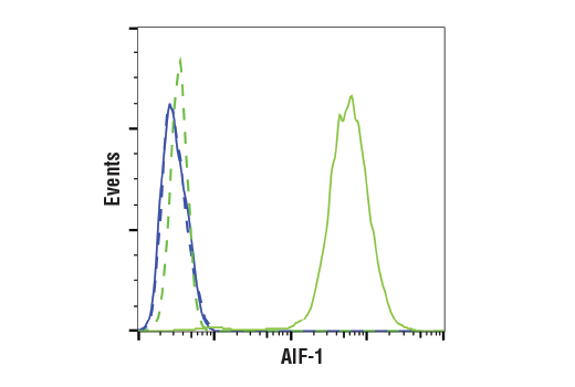
Flow cytometric analysis of SH-SY5Y cells (blue, negative) and THP-1 cells (green, positive) using Iba1/AIF-1 (E4O4W) XP® Rabbit mAb (solid lines) or a concentration-matched Rabbit (DA1E) mAb IgG XP® Isotype Control #3900 (dashed lines). Anti-rabbit IgG (H+L), F(ab')2 Fragment (Alexa Fluor® 488 Conjugate) #4412 was used as a secondary antibody.
Immunoprecipitation of HS1 protein from We-HI231 cell extracts. Lane 1 is HS1 (D5A9) XP® Rabbit mAb, lane 2 is Rabbit (DA1E) mAb IgG XP® Isotype Control #3900, and lane 3 is 10% input. Western blot analysis was performed using HS1 (D5A9) XP® Rabbit mAb #3892. Anti-rabbit IgG, HRP-linked Antibody #7074 was used as a secondary antibody.
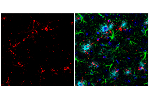
Confocal immunofluorescent analysis of mouse subiculum using CD68 (E3O7V) Rabbit mAb (red). Free secondary binding sites were then blocked with Rabbit (DA1E) mAb IgG XP® Isotype Control #3900 prior to colabeling with GFAP (GA5) Mouse mAb (Alexa Fluor® 488 Conjugate) #3655 (right, green), β-Amyloid (D54D2) XP® Rabbit mAb (Alexa Fluor® 647 Conjugate) #42284 (right, cyan pseudocolor), and DAPI #4083 (right, blue).
Confocal immunofluorescent analysis of mouse subiculum from the 5XFAD mouse model of Alzheimer's disease using CD68 (E3O7V) Rabbit mAb (red). Free secondary binding sites were then blocked with Rabbit (DA1E) mAb IgG XP® Isotype Control #3900 prior to colabeling with GFAP (GA5) Mouse mAb (Alexa Fluor® 488 Conjugate) #3655 (right, green), β-Amyloid (D54D2) XP® Rabbit mAb (Alexa Fluor® 647 Conjugate) #42284 (right, cyan pseudocolor), and DAPI #4083 (right, blue).