


 全部商品分类
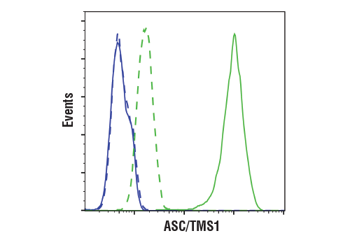


 下载产品说明书 下载SDS
 用小程序,查商品更便捷


 收藏
收藏
 对比
对比 咨询
咨询
The Mouse Reactive Pyroptosis Antibody Sampler Kit provides an economical means of detecting proteins that are used as readouts for pyroptosis. The kit includes enough antibodies to perform two western blot experiments with each primary antibody.
参考图片
Flow cytometric analysis of Raw264.7 cells (blue) and J774A.1 cells (green) using ASC/TMS1 (D2W8U) Rabbit mAb (solid lines) or a concentration-matched Rabbit (DA1E) mAb IgG XP® Isotype Control #3900 (dashed lines). Anti-rabbit IgG (H+L), F(ab')2 Fragment (Alexa Fluor® 488 Conjugate) #4412 was used as a secondary antibody.
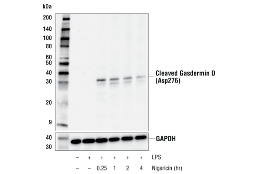
Western blot analysis of extracts from mouse bone marrow derived macrophages (mBMDM), untreated (-) or treated with Lipopolysaccharides (LPS) #14011 (50 ng/ml, 4 hr; +) followed by Nigericin (sodium salt) #66419 (15 μM, indicated times; +), using Cleaved Gasdermin D (Asp276) (E3E3P) Rabbit mAb (upper) or GAPDH (D16H11) XP® Rabbit mAb #5174 (lower).
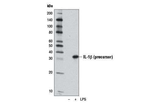
Western blot analysis of extracts from Raw 264.7 cells, untreated (-) or LPS-treated (1 μg/ml, overnight; +), using IL-1β (D3H1Z) Rabbit mAb.
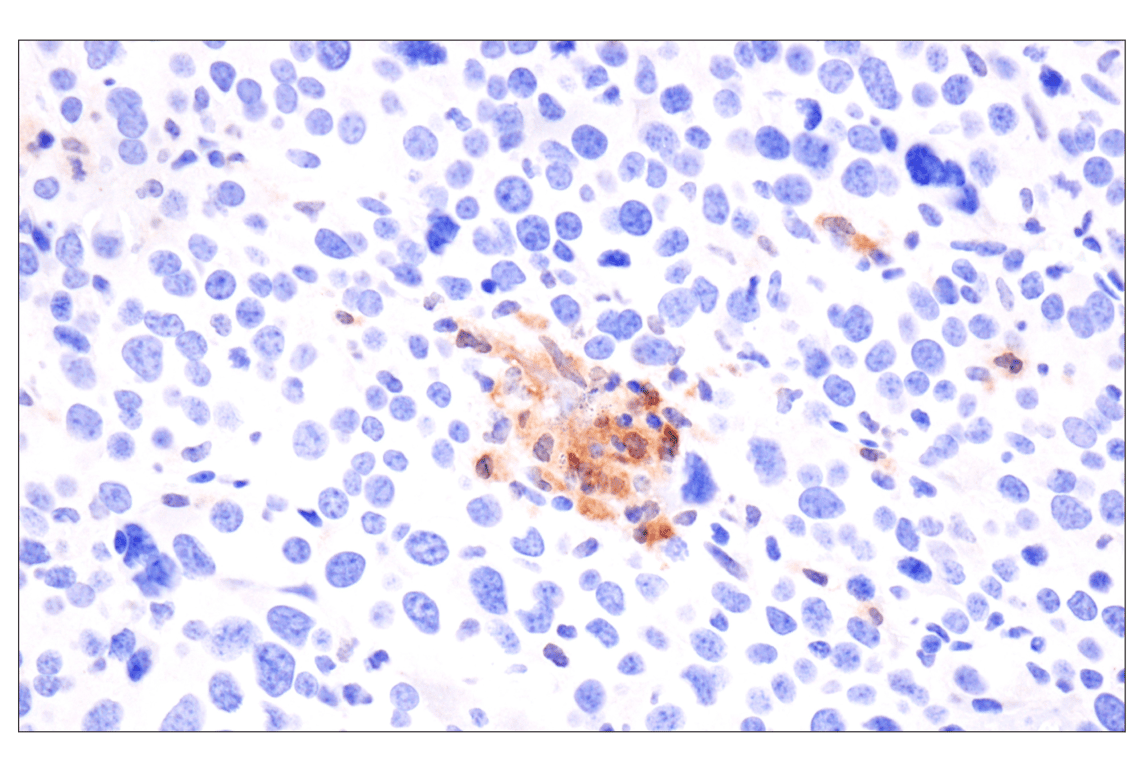
Immunohistochemical analysis of paraffin-embedded Renca syngeneic tumor using IL-1β (D3H1Z) Rabbit mAb.
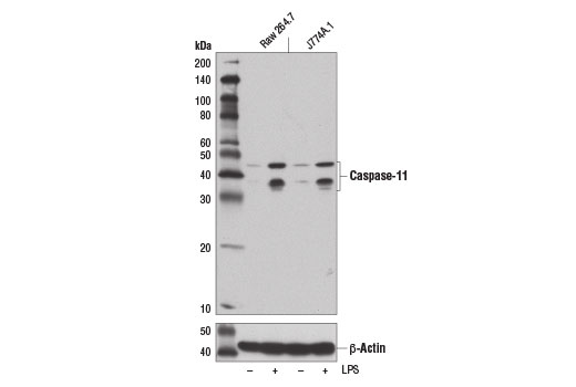
Western blot analysis of extracts from Raw 264.7 or J774.1 cells, untreated (-) or treated with LPS (1 μg/ml, overnight; +) using Caspase-11 (17D9) Rat mAb (upper) or β-Actin (D6A8) Rabbit mAb #8457 (lower).
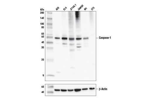
Western blot analysis of extracts from various cell lines using Caspase-1 (E2Z1C) Rabbit mAb (upper), or β-Actin (D6A8) Rabbit mAb #8457 (lower).
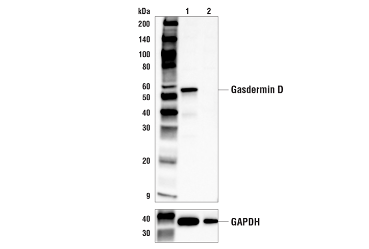
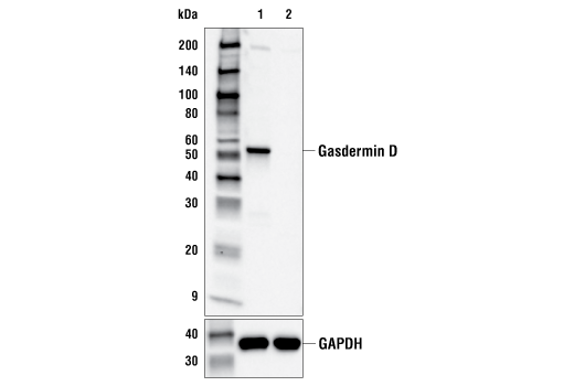
Western blot analysis of extracts from control mouse bone marrow derived macrophages (mBMDM; lane 1) or mBMDM from Gasdermin D knockout mice (lane 2) using Gasdermin D (E9S1X) Rabbit mAb (upper) or GAPDH (D16H11) XP® Rabbit mAb #5174 (lower). The absence of signal in the Gasdermin D knockout cells confirms the specificity of the antibody for Gasdermin D. The mBMDM from Gasdermin D knockout mice were kindly provided by Dr. Douglas Golenbock, M.D., University of Massachusetts Medical School, Worcester, MA.
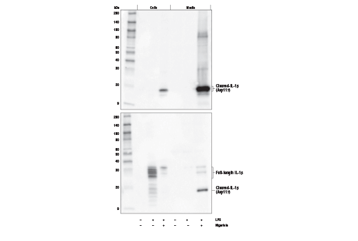
Western blot analysis of cell extracts and media from mouse bone marrow derived macrophages (mBMDM), untreated (-), or treated (+) with combinations of LPS #14011 (50 ng/ml, 4 hr) followed by nigericin (15 μM, 45 min) using Cleaved-IL-1β (Asp117) (D7V2A) Rabbit mAb (upper) or total IL-1β (E3H1Z) Rabbit mAb #12507 (lower).
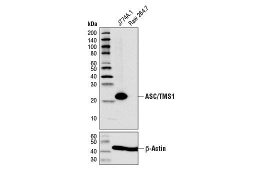
Western blot analysis of extracts from J774A.1 and Raw 264.7 cells using ASC/TMS1 (D2W8U) Rabbit mAb (upper) or β-Actin (D6A8) Rabbit mAb #8457 (lower).
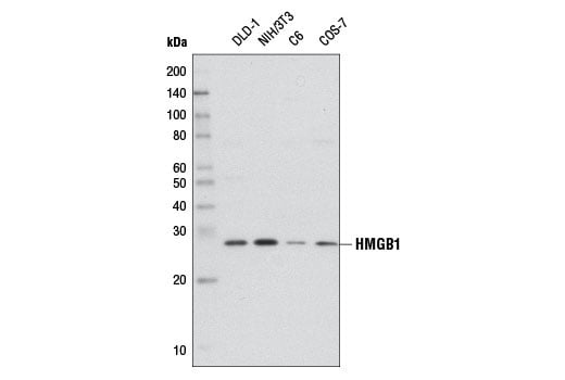
Western blot analysis of extracts from various cell lines using HMGB1 (D3E5) Rabbit mAb.
After the primary antibody is bound to the target protein, a complex with HRP-linked secondary antibody is formed. The LumiGLO® is added and emits light during enzyme catalyzed decomposition.
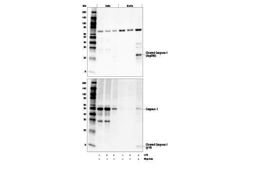
Western blot analysis of cell extracts from the cells or media from mouse bone marrow derived macrophages (mBMDM), untreated (-) or treated with Lipopolysaccharides (LPS) #14011 (50 ng/ml, 4 hr) followed by Nigericin (15 μM, 45 min) (+), using Cleaved Caspase-1 (Asp296) (E2G2I) Rabbit mAb (upper), or Caspase-1 (E2Z1C) Rabbit mAb (lower).
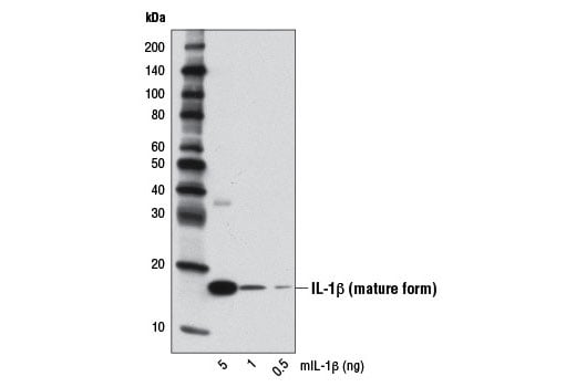
Western blot analysis of Mouse Interleukin-1β (mIL-1β) #5204 using IL-1β (D3H1Z) Rabbit mAb.
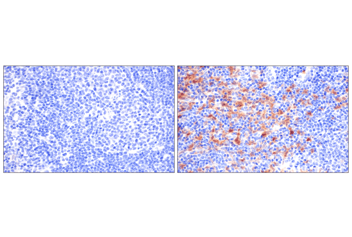
Immunohistochemical analysis of paraffin-embedded mouse spleen, untreated (left) or treated with Lipopolysaccharides (LPS) #14011 (right), using IL-1β (D3H1Z) Rabbit mAb.
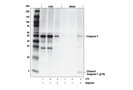
Western blot analysis of cell extracts from the cells or media from mouse bone marrow derived macrophages (mBMDM), untreated (-) or treated with Lipopolysaccharides (LPS) #14011 (50 ng/ml, 4hr) followed by Nigericin (15 μM, 45 min) (+) using Caspase-1 (E2Z1C) Rabbit mAb.
Western blot analysis of extracts from control PC-3 cells (lane 1) or Gasdermin D knockout PC-3 cells (lane 2) using Gasdermin D (E9S1X) Rabbit mAb (upper) or GAPDH (D16H11) XP® Rabbit mAb #5174 (lower). The absence of signal in the Gasdermin D knockout PC-3 cells confirms specificity of the antibody for Gasdermin D.
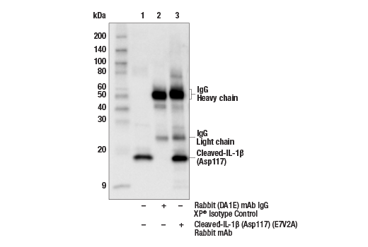
Immunoprecipitation of Cleaved-IL-1β (Asp117) from extracts of media from mouse bone marrow derived macrophages (mBMDM) treated with LPS #14011 (50 ng/ml, 4 hr) followed by nigericin (15 μM, 45 min). Lane 1 is 10% input, lane 2 is Rabbit (DA1E) mAb IgG XP® Isotype Control #3900, and lane 3 is Cleaved-IL-1β (Asp117) (E7V2A) Rabbit mAb. Western blot was performed using Cleaved-IL-1β (Asp117) (E7V2A) Rabbit mAb. Anti-Rabbit IgG, HRP-linked Antibody #7074 was used as a secondary antibody.
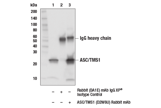
Immunoprecipitation of ASC/TMS1 from J774A.1 cell extracts. Lane 1 is 10% input, lane 2 is Rabbit (DA1E) mAb IgG XP® Isotype Control #3900, and lane 3 is ASC (D2W8U) Rabbit mAb. Western blot analysis was performed using ASC/TMS1 (D2W8U) Rabbit mAb.
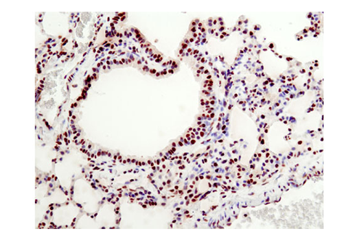
Immunohistochemical analysis of paraffin-embedded mouse lung using HMGB1 (D3E5) Rabbit mAb.
Western blot analysis of extracts from the media of mouse bone marrow derived macrophages (mBMDM), untreated (-) or treated with Lipopolysaccharides (LPS) #14011 (50 ng/ml, 4 hr; +) followed by Nigericin (15 μM, 45 min; +), using HMGB1 (D3E5) Rabbit mAb.
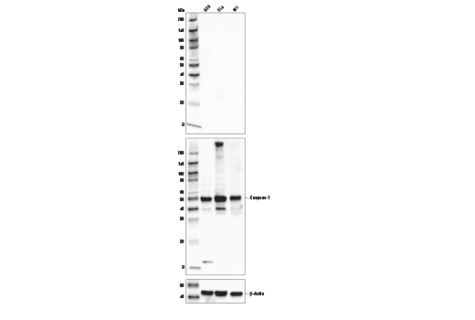
Western blot of A20, EL4, and M1 cell lines using Cleaved Caspase-1 (Asp296) (E2G2I) Rabbit mAb (upper), Caspase-1 (E2Z1C) Rabbit mAb (middle), or β-Actin (D6A8) Rabbit mAb #8457 (lower). The lack of staining in these cell lines using Cleaved Caspase-1 (Asp296) (E2G2I) Rabbit mAb demonstrates that it does not cross-react with full-length caspase-1.
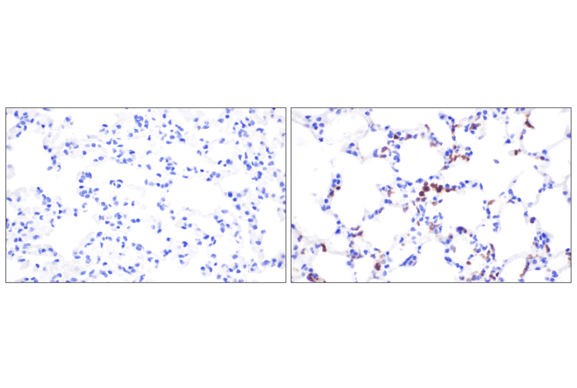
Immunohistochemical analysis of paraffin-embedded mouse lung, untreated (left) or treated with Lipopolysaccharides (LPS) #14011 (right), using IL-1β (D3H1Z) Rabbit mAb.
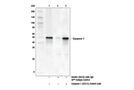
Immunoprecipitation of Caspase-1 from EL4 cell extracts. Lane 1 is 10% input, lane 2 is Rabbit (DA1E) mAb IgG XP® Isotype Control #3900, and lane 3 Caspase-1 (E2Z1C) Rabbit mAb. Western blot was performed using Caspase-1 (E2Z1C) Rabbit mAb. Mouse Anti-rabbbit IgG (Conformation Specific) (L27A9) mAb (HRP Conjugate) #5127 was used as a secondary antibody.
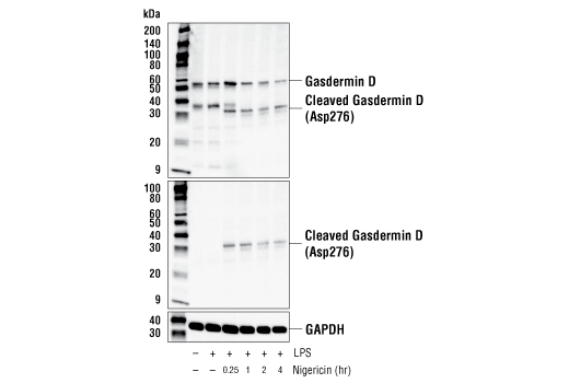
Western blot analysis of extracts from mouse bone marrow derived macrophages (mBMDM), untreated (-) or treated with Lipopolysaccharides (LPS) #14011 (50 ng/ml, 4 hr; +) followed by Nigericin (sodium salt) #66419 (15 μM, indicated times), using Gasdermin D (E9S1X) Rabbit mAb (upper), Cleaved Gasdermin D (Asp276) (E3E3P) Rabbit mAb #10137 (middle), or GAPDH (D16H11) XP® Rabbit mAb #5174 (lower).
Immunohistochemical analysis of paraffin-embedded J774A.1 cell pellet (left, positive) or RAW 264.7 cell pellet (right, negative) using ASC/TMS1 (D2W8U) Rabbit mAb.
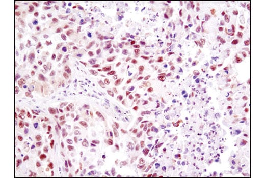
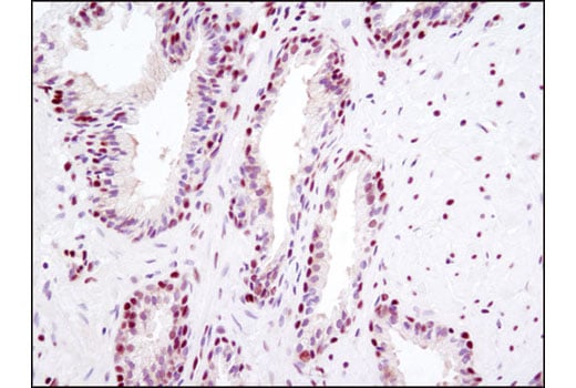
Immunohistochemical analysis of paraffin-embedded human colon carcioma using HMGB1 (D3E5) Rabbit mAb.
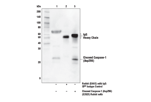
Immunoprecipitation of Cleaved Caspase-1 (Asp296) from extracts of acetone precipitated media from mouse bone marrow derived macrophages treated with Lipopolysaccharides (LPS) #14011 (50ng/ml, 4hr) followed by Nigericin (15 μM, 45 min). Lane 1 is 10% input, lane 2 is Rabbit (DA1E) mAb IgG XP® Isotype Control #3900, and lane 3 is Cleaved Caspase-1 (Asp296) (E2G2I) Rabbit mAb. Western blot analysis was performed using Cleaved Caspase-1 (Asp296) (E2G2I) Rabbit mAb. Anti-rabbit IgG, HRP-linked Antibody #7074 was used as a secondary antibody.
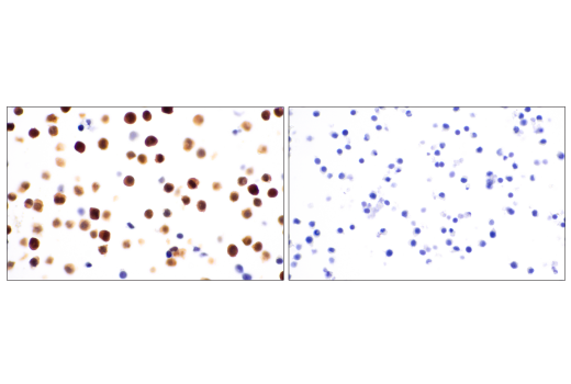
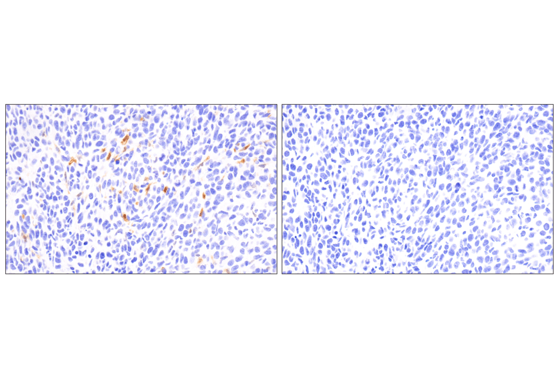
Immunohistochemical analysis of paraffin-embedded CT26.WT syngeneic tumor using IL-1β (D3H1Z) Rabbit mAb (left) compared to concentration-matched Rabbit (DA1E) mAb IgG XP® Isotype Control #3900 (right).
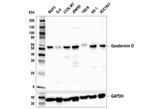
Western blot analysis of extracts from various cell lines using Gasdermin D (E9S1X) Rabbit mAb (upper) or GAPDH (D16H11) XP® Rabbit mAb #5174 (lower).
Immunohistochemical analysis of paraffin-embedded mouse forestomach using ASC/TMS1 (D2W8U) Rabbit mAb.
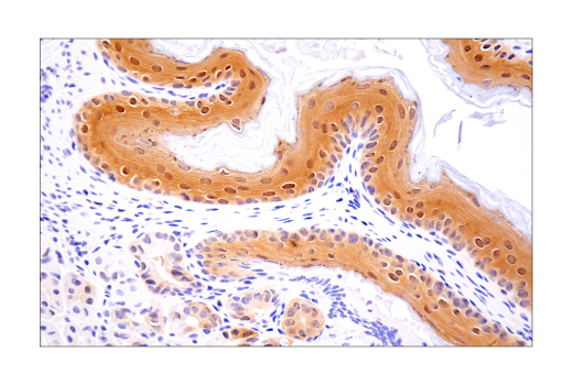
Immunohistochemical analysis of paraffin-embedded human lung carcioma using HMGB1 (D3E5) Rabbit mAb.
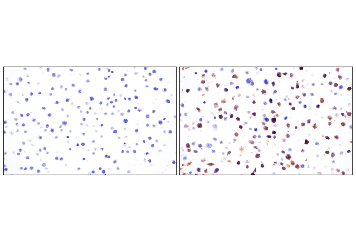
Immunohistochemical analysis of paraffin-embedded RAW 264.7 cell pellets, untreated (left, negative) or treated with Lipopolysaccharides (LPS) #14011 (100 ng/ml, 7 hr), (positive, right), using IL-1β (D3H1Z) Rabbit mAb.
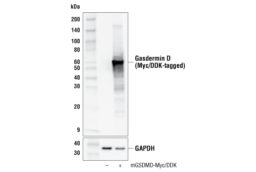
Western blot analysis of extracts from 293T cells, untransfected (-) or transfected with a construct expressing Myc/DDK-tagged full-length mouse Gasdermin D protein (mGSDMD-Myc/DDK; +), using Gasdermin D (E9S1X) Rabbit mAb (upper) or GAPDH (D16H11) XP® Rabbit mAb #5174 (lower).
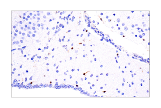
Immunohistochemical analysis of paraffin-embedded mouse brain using ASC/TMS1 (D2W8U) Rabbit mAb.
Immunohistochemical analysis of paraffin-embedded human prostate carcioma using HMGB1 (D3E5) Rabbit mAb.
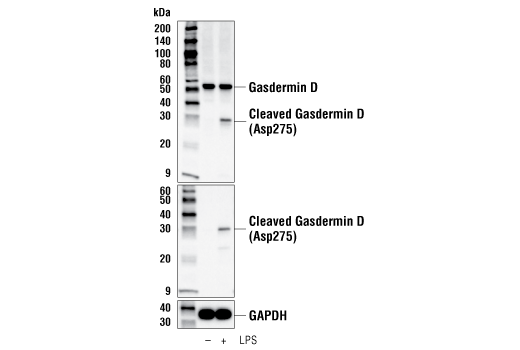
Western blot analysis of extracts from THP-1 cells, differentiated with TPA (12-O-Tetradecanoylphorbol-13-Acetate) #4174 (50 ng/ml, overnight) and then treated with Lipopolysaccharides (LPS) #14011 (5 μg/ml, 6 hr), using Gasdermin D (E9S1X) Rabbit mAb (upper), Cleaved Gasdermin D (Asp275) (E7H9G) Rabbit mAb #36425 (middle), or GAPDH (D16H11) XP® Rabbit mAb #5174 (lower).
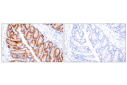
Immunohistochemical analysis of paraffin-embedded mouse colon using ASC/TMS1 (D2W8U) Rabbit mAb (left) compared to concentration-matched Rabbit (DA1E) mAb IgG XP® Isotype Control #3900 (right).
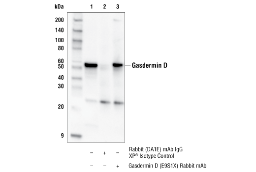
Immunoprecipitation of Gasdermin D protein from J774A.1 cell extracts. Lane 1 is 10% input, lane 2 is Rabbit (DA1E) mAb IgG XP® Isotype Control #3900, and lane 3 is Gasdermin D (E9S1X) Rabbit mAb. Western blot analysis was performed using Gasdermin D (E9S1X) Rabbit mAb. Mouse Anti-rabbit IgG (Conformation Specific) (L27A9) mAb (HRP Conjugate) #5127 was used as a secondary antibody.
Immunohistochemical analysis of paraffin-embedded mouse thymus using ASC/TMS1 (D2W8U) Rabbit mAb.
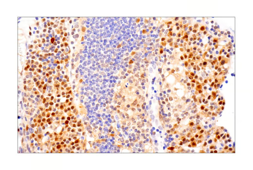
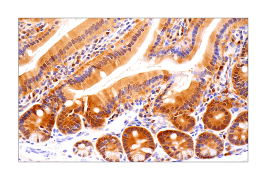
Immunohistochemical analysis of paraffin-embedded mouse small intestine using ASC/TMS1 (D2W8U) Rabbit mAb.
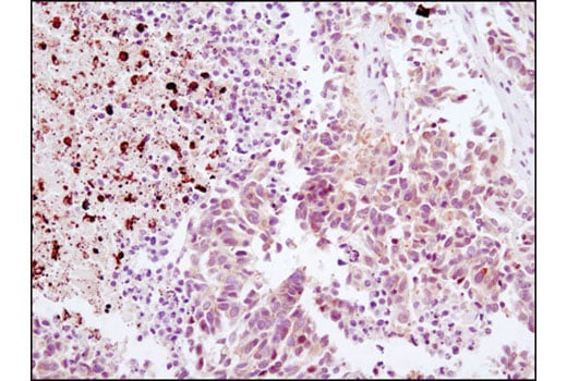
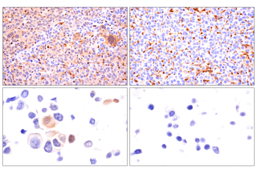
Immunohistochemical analysis of paraffin-embedded Renca syngeneic tumor (top left), 4T1 syngeneic mammary tumor (top right), Renca cell pellet (bottom left), and 4T1 cell pellet (bottom right) using ASC/TMS1 (D2W8U) Rabbit mAb. Both tumors show staining of infiltrating immune cells. Note the presence of staining in the Renca tumor cells and the lack of staining in the 4T1 tumor cells consistent with staining results on corresponding cell pellets.
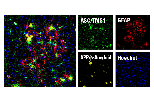
Confocal immunofluorescent analysis of mouse Tg2576 brain which overexpresses mutant human APP695. Sections were first labeled with ASC/TMS1 (D2W8U) Rabbit mAb #67824 (green) and APP/β-Amyloid (NAB228) Mouse mAb #2450 (yellow). After blocking free secondary binding sites with Mouse (G3A1) mAb IgG1 Isotype Control #5415, sections were incubated with GFAP (GA5) Mouse mAb (Alexa Fluor® 647 Conjugate) #3657 (red). Nuclei were labeled with Hoechst 33342 #4082 (blue).
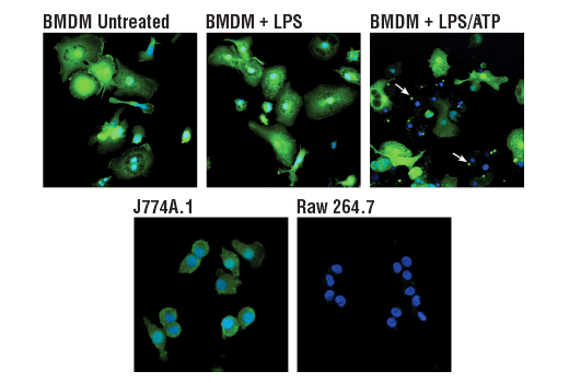
Confocal immunofluorescent analysis of mouse primary bone marrow-derived macrophages (BMDMs) either untreated (upper left) or treated with LPS (50 ng/ml, 4 hr, middle) or LPS followed by ATP (5 mM, 45 min, upper right), and J774A.1 (lower left) or Raw 264.7 (lower right) cells, using ASC/TMS1 (D2W8U) Rabbit mAb (green). Blue pseudocolor = DRAQ5® #4084 (fluorescent DNA dye). Note the translocation of ASC to inflammasomes following stimulation with LPS and ATP (white arrows).