


 全部商品分类


1/2
PE-Cy5 Mouse Anti-Human CD206(19.2)

品牌: BD Pharmingen
 下载产品说明书
 用小程序,查商品更便捷


 收藏
收藏
 对比
对比 咨询
咨询反应种属:
Human (QC Testing)
来源宿主:
Mouse IgG1, κ
产品介绍
产品使用步骤推荐 产品介绍
产品信息
荧光素标记
抗原名称
CD206

宿主
Mouse IgG1, κ
免疫原
Human Placental Macrophage Mannose Receptor
简单描述
The 19.2 monoclonal antibody specifically binds to CD206 which is also known as the macrophage mannose receptor (MMR) or C-type lectin domain family 13 member D (CLEC13D). CD206 is a type I transmembrane glycoprotein of approximately 162 kDa which binds to glycoconjugates containing mannose, fucose, or N-acetylglucosamine. These carbohydrates are present on the surface of many microorganisms and enable the macrophage to bind to microorganisms through the MMR and internalize them during the process of phagocytosis. CD206 is expressed on human macrophages, endothelial cells, and cultured dendritic cells. It is not detected on resting monocytes. Mannose receptor expression is upregulated on peripheral blood mononuclear cells following 3 day incubation with GM-CSF.
This antibody is routinely by flow cytometric analysis. Other applications were tested at BD Biosciences Pharmingen during antibody development only or reported in the literature.
商品描述
The 19.2 monoclonal antibody specifically binds to CD206 which is also known as the macrophage mannose receptor (MMR) or C-type lectin domain family 13 member D (CLEC13D). CD206 is a type I transmembrane glycoprotein of approximately 162 kDa which binds to glycoconjugates containing mannose, fucose, or N-acetylglucosamine. These carbohydrates are present on the surface of many microorganisms and enable the macrophage to bind to microorganisms through the MMR and internalize them during the process of phagocytosis. CD206 is expressed on human macrophages, endothelial cells, and cultured dendritic cells. It is not detected on resting monocytes. Mannose receptor expression is upregulated on peripheral blood mononuclear cells following 3 day incubation with GM-CSF.
This antibody is routinely by flow cytometric analysis. Other applications were tested at BD Biosciences Pharmingen during antibody development only or reported in the literature.
同种型
Mouse IgG1, κ
克隆号
19.2
产品详情
PE-Cy5
PE-Cy5 dye is a part of the BD PE family of dyes. This tandem fluorochrome is comprised of a R-Phycoerythrin (PE) donor that has excitation maxima (Ex Max) of 496 nm and 566 nm and an acceptor dye, Cy™5, with an emission maximum (Em Max) at 670-nm. PE designed to be excited by the Blue (488-nm), Green (532-nm) and yellow-green (561-nm) lasers and detected using an optical filter centered near 670-nm (e.g., a 670/20-nm bandpass filter). The donor dye can be excited by the Blue (488-nm), Green (532-nm) and yellow-green (561-nm) lasers and the acceptor dye can be excited by the Red (627-640-nm) laser resulting in cross-laser excitation and fluorescence spillover. Please ensure that your instrument’s configurations (lasers and optical filters) are appropriate for this dye.
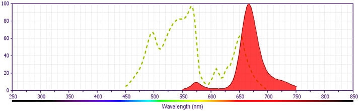
PE-Cy5
Yellow-Green 488 nm, 532 nm, 561 nm
496 nm, 566 nm
670 nm
应用
实验应用
Flow cytometry (Routinely Tested)
推荐用量
20 µl
反应种属
Human (QC Testing)
背景
别名
Macrophage mannose receptor; MMR; MRC1; CLEC13D
制备和贮存
存储溶液
Aqueous buffered solution containing BSA and ≤0.09% sodium azide.
保存方式
Aqueous buffered solution containing BSA and ≤0.09% sodium azide.
文献
文献
研发参考(2)
1. Mason D. David Mason .. et al., ed. Leucocyte typing VII : white cell differentiation antigens : proceedings of the Seventh International Workshop and Conference held in Harrogate, United Kingdom. Oxford: Oxford University Press; 2002.
2. Pontow SE, Kery V, Stahl PD. Mannose receptor. Int Rev Cytol. 1992; 137(B):221-244. (Clone-specific).
数据库链接
Entrez-Gene ID
4360
参考图片
Profile of GM-CSF-stimulated (3 days) peripheral blood mononuclear cells analyzed by flow cytometry.
声明 :本官网所有报价均为常温或者蓝冰运输价格,如有产品需要干冰运输,需另外加收干冰运输费。