Recombinant Mouse CD206 Carbohydrate recognition domains 4-7/Fc Fusion Protein
简单描述
The MR5D3 monoclonal antibody specifically binds to CD206 which is also known as the Macrophage mannose receptor (MMR, MR) or Mannose receptor, C type 1 (Mrc1). CD206 is a type I transmembrane glycoprotein of approximately 175 kDa that belongs to the C-type lectin superfamily. It is expressed at the cell surface and intracellularly by macrophages, Langerhans cells, dendritic cells, and endothelial cells within hepatic and lymphoid tissues. This pattern recognition receptor binds to endogenous and microbial glycoconjugates containing mannose, fucose, or N-acetylglucosamine through its C-type lectin-like carbohydrate recognition domains (CRD). CD206 also contains a cysteine-rich domain that enables binding to sulfated carbohydrate antigens. This receptor enables macrophages and other specialized cells to maintain tissue homeostasis as well as to internalize microbes or their components by phagocytosis or endocytosis. CD206 thus plays important roles in mediating innate immunity, e.g., enabling phagocytosis, as well as in processing and presenting antigens for the generation and expression of adaptive immunity. Moreover, CD206 has been associated with leucocyte homing and cancer cell metastasis.
商品描述
MR5D3
The MR5D3 monoclonal antibody specifically binds to CD206 which is also known as the Macrophage mannose receptor (MMR, MR) or Mannose receptor, C type 1 (Mrc1). CD206 is a type I transmembrane glycoprotein of approximately 175 kDa that belongs to the C-type lectin superfamily. It is expressed at the cell surface and intracellularly by macrophages, Langerhans cells, dendritic cells, and endothelial cells within hepatic and lymphoid tissues. This pattern recognition receptor binds to endogenous and microbial glycoconjugates containing mannose, fucose, or N-acetylglucosamine through its C-type lectin-like carbohydrate recognition domains (CRD). CD206 also contains a cysteine-rich domain that enables binding to sulfated carbohydrate antigens. This receptor enables macrophages and other specialized cells to maintain tissue homeostasis as well as to internalize microbes or their components by phagocytosis or endocytosis. CD206 thus plays important roles in mediating innate immunity, e.g., enabling phagocytosis, as well as in processing and presenting antigens for the generation and expression of adaptive immunity. Moreover, CD206 has been associated with leucocyte homing and cancer cell metastasis.
同种型
Rat F344, also known as Fischer, CDF IgG2a
克隆号
克隆 MR5D3 (RUO)
浓度
0.2 mg/ml
产品详情
Alexa Fluor™ 647
Alexa Fluor™ 647 Dye is part of the BD red family of dyes. This is a small organic fluorochrome with an excitation maximum (Ex Max) at 653-nm and an emission maximum (Em Max) at 669-nm. Alexa Fluor 647 is designed to be excited by the Red laser (627-640 nm) and detected using an optical filter centered near 520-nm (e.g., a 660/20 nm bandpass filter). Please ensure that your instrument’s configurations (lasers and optical filters) are appropriate for this dye.
研发参考(5)
1. Akbarshahi H, Menzel M, Posaric Bauden M, Rosendahl A, Andersson R. Enrichment of murine CD68+ CCR2+ and CD68+ CD206+ lung macrophages in acute pancreatitis-associated acute lung injury. PLoS ONE. 2012; 7(10):e42654. (Biology).
2. Burgdorf S, Lukacs-Kornek V, Kurts C. The mannose receptor mediates uptake of soluble but not of cell-associated antigen for cross-presentation. J Immunol. 2006; 176(11):6770-6776. (Biology).
3. Marttila-Ichihara F, Turja R, Miiluniemi M, et al. Macrophage mannose receptor on lymphatics controls cell trafficking. Blood. 2008; 112(1):64-72. (Clone-specific: Immunofluorescence, Immunohistochemistry).
4. McKenzie EJ, Taylor PR, Stillion RJ, et al. J Immunol. 2007; 178(8):4975-4983. (Clone-specific: Flow cytometry).
5. Zamze S, Martinez-Pomares L, Jones H, et al. Recognition of bacterial capsular polysaccharides and lipopolysaccharides by the macrophage mannose receptor. J Biol Chem. 2002; 277(44):41613-41623. (Immunogen: Dot Blot, ELISA, Flow cytometry, Immunoaffinity chromatography, Immunohistochemistry, Immunoprecipitation).
参考图片
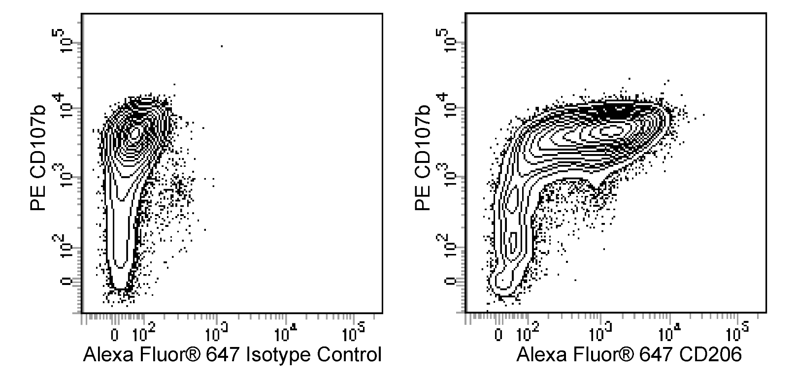
Two-color flow cytometric analysis of CD206 expression on mouse peritoneal exudate cells. Thioglycollate-elicited BALB/c mouse peritoneal exudate cells (PEC) were fixed and permeabilized using the BD Pharmingen™ Transcription Factor Buffer Set (Cat. No. 562574/562725). The leucocytes were then stained with PE Rat Anti-Mouse CD107b (Mac-3) antibody (Cat. No. 553324) and either Alexa Fluor® 647 Rat IgG2a, κ Isotype Control (Cat. No. 565250; Left Panel) or Alexa Fluor® 647 Rat Anti-Mouse CD206 antibody (Cat. No. 565250; Right Panel). Two-color flow cytometric contour plots showing the correlated expression of CD206 (or Ig Isotype control staining) versus CD107b were derived from gated events with the forward and side-light scattering characteristics of intact PEC. Flow cytometric analysis was performed using a BD FACSCanto™ II Flow Cytometer System.
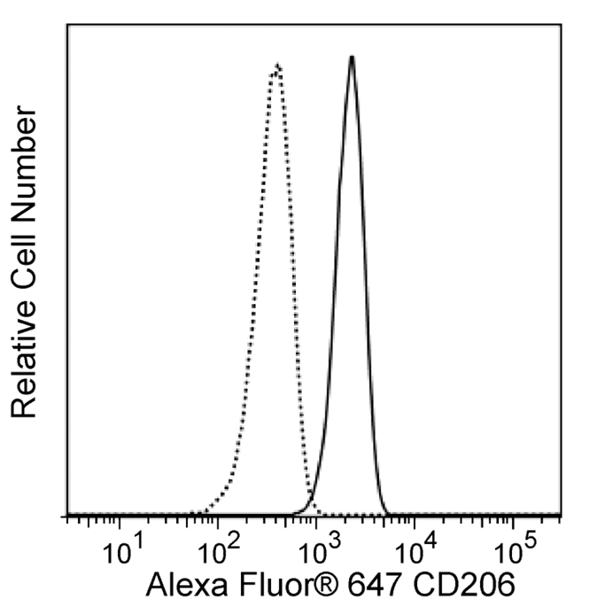
Flow cytometric analysis of CD206 expression by J774A cells. Cells from the J774A (Mouse macrophage, ATCC TIB-67) cell line were similarly fixed and permeabilized, stained with either Alexa Fluor® 647 Rat IgG2a, κ Isotype Control (dashed line histogram) or Alexa Fluor® 647 Rat Anti-Mouse CD206 antibody (solid line histogram), and analzyed by flow cytometry. The fluorescence histogram showing CD206 expression (or Ig Isotype control staining) was derived from gated events with the forward and side light-scatter characteristics of intact J774A cells.


 全部商品分类


 下载产品说明书
 用小程序,查商品更便捷


 收藏
收藏
 对比
对比 咨询
咨询