


 全部商品分类


BD Horizon™ BV650 Rat Anti-Mouse CD86


 下载产品说明书 下载SDS
 用小程序,查商品更便捷


 收藏
收藏
 对比
对比 咨询
咨询

参考图片
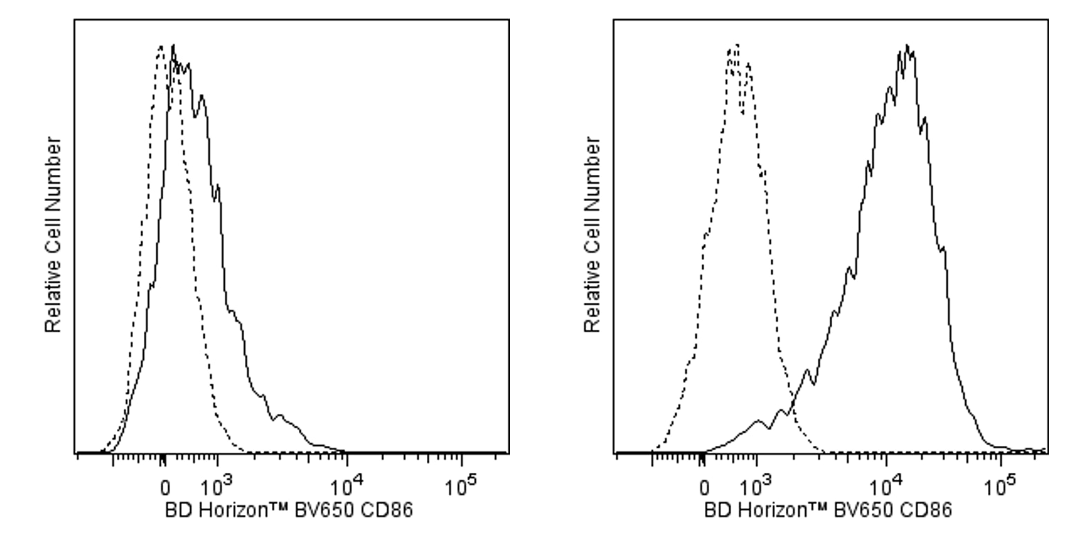
Flow cytometric analysis of CD86 expression on resting or activated mouse splenocytes. Freshly isolated (Left Panel) or 72-hour lipopolysaccharide-stimulated (Right Panel) mouse splenic leucocytes were pretreated with Purified Rat Anti-Mouse CD16/CD32 antibody (Mouse BD Fc Block™) (Cat. No. 553141/553142). The cells were then stained with either BD Horizon™ BV650 Rat IgG2a, κ Isotype Control (Cat. No. 563236; dashed line histograms) or BD Horizon BV650 Rat Anti-Mouse CD86 antibody (Cat. No. 564200; solid line histograms). The fluorescence histograms were derived from gated events with the forward and side light-scatter characteristics of viable resting (Left Panel) or activated (Right Panel) lymphocytes. Flow cytometric analysis was performed using a BD™ LSR II Flow Cytometer System.
Flow cytometric analysis of CD86 expression on resting or activated mouse splenocytes. Freshly isolated (Left Panel) or 72-hour lipopolysaccharide-stimulated (Right Panel) mouse splenic leucocytes were pretreated with Purified Rat Anti-Mouse CD16/CD32 antibody (Mouse BD Fc Block™) (Cat. No. 553141/553142). The cells were then stained with either BD Horizon™ BV650 Rat IgG2a, κ Isotype Control (Cat. No. 563236; dashed line histograms) or BD Horizon BV650 Rat Anti-Mouse CD86 antibody (Cat. No. 564200; solid line histograms). The fluorescence histograms were derived from gated events with the forward and side light-scatter characteristics of viable resting (Left Panel) or activated (Right Panel) lymphocytes. Flow cytometric analysis was performed using a BD™ LSR II Flow Cytometer System.