


 下载产品说明书
 用小程序,查商品更便捷


 收藏
收藏
 对比
对比 咨询
咨询
Specificity/Sensitivity
参考图片
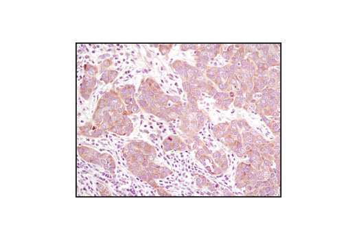
Immunohistochemical analysis of paraffin-embedded human lymphoma using RagC (D31G9) XP® Rabbit mAb in the presence of control peptide (left) or antigen-specific peptide (right).
After the primary antibody is bound to the target protein, a complex with HRP-linked secondary antibody is formed. The LumiGLO* is added and emits light during enzyme catalyzed decomposition.
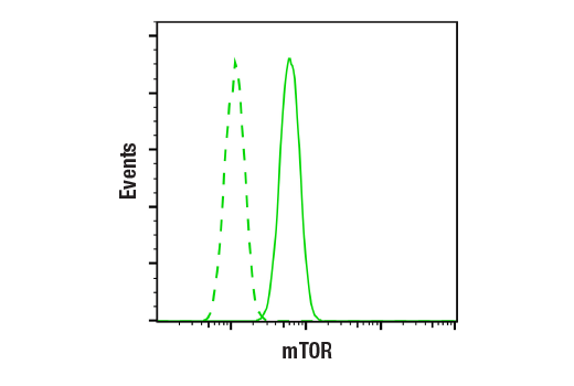
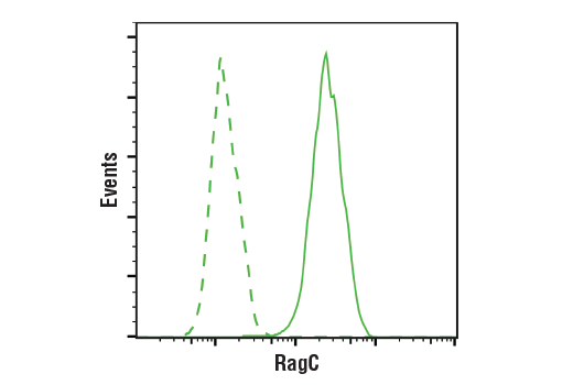
Flow cytometric analysis of 293 cells using mTOR (7C10) Rabbit mAb (blue) compared to a nonspecific negative control antibody (red).
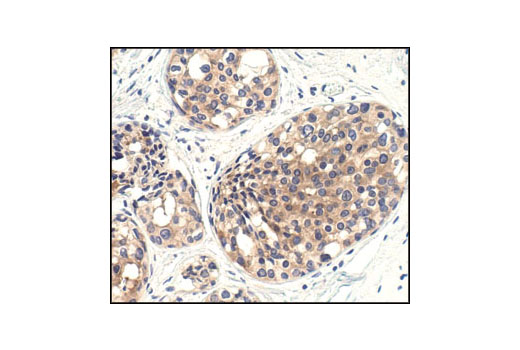
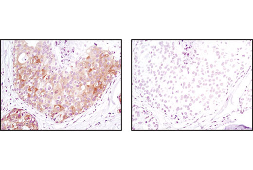
Immunohistochemical analysis of paraffin-embedded human breast carcinoma, showing cytoplasmic localization using mTOR (7C10) Rabbit mAb.
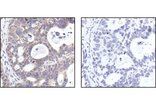
Immunohistochemical analysis of paraffin-embedded human lung carcinoma, using mTOR (7C10) Rabbit mAb in the presence of control peptide (left) or mTOR Blocking Peptide #1072 (right).
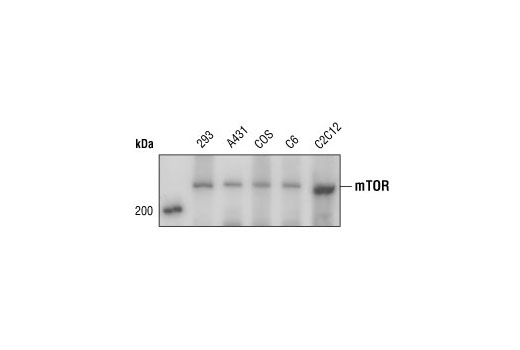
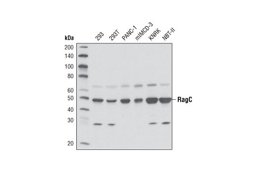
Western blot analysis of extracts from 293, A431, COS, C6, and C2C12 cells, using mTOR (7C10) Rabbit mAb.
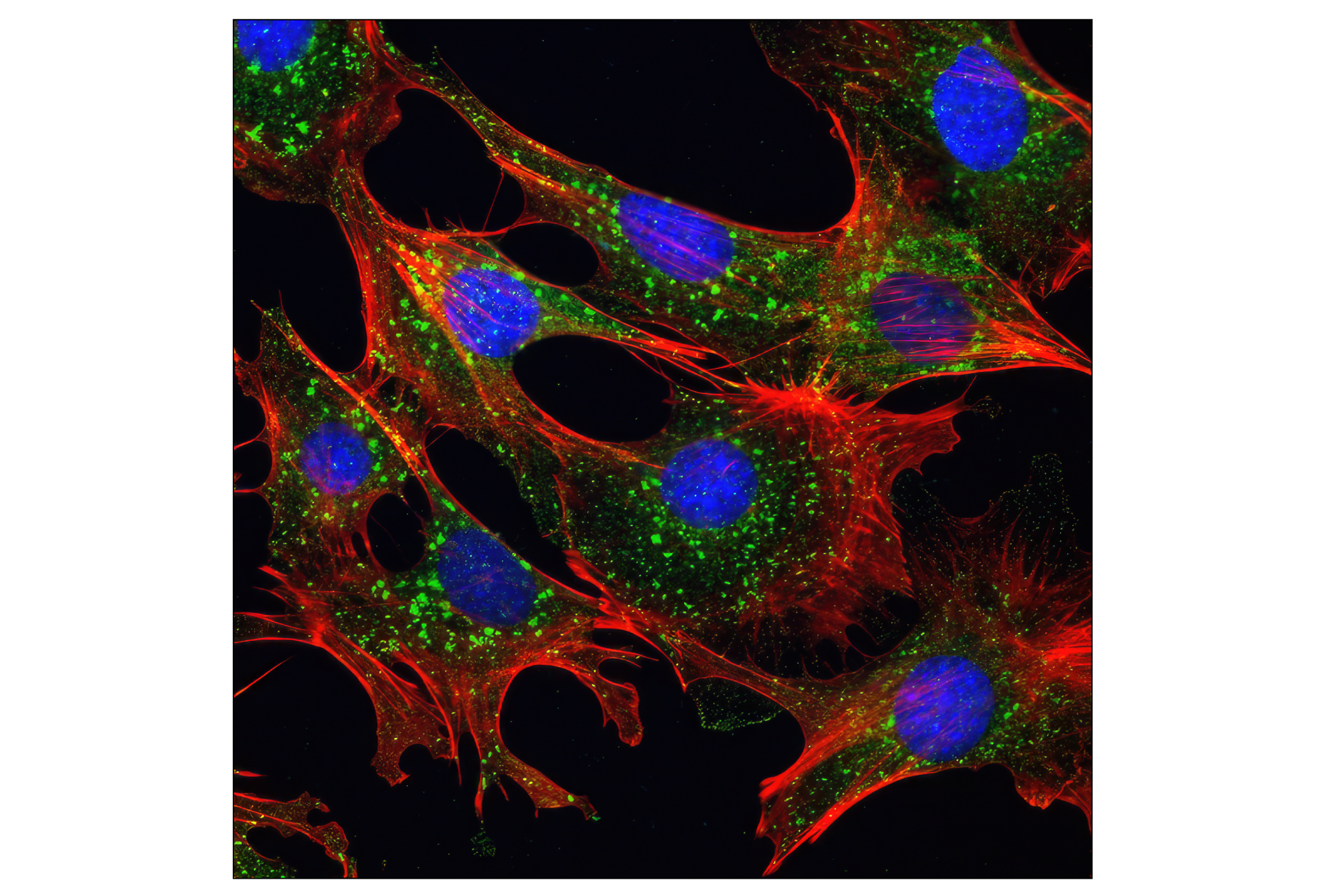
Confocal immunofluorescent analysis of mouse embryonic fibroblast (MEF) cells using mTOR (7C10) Rabbit mAb (green). Actin filaments were labeled with DY-554 phalloidin (red). Blue pseudocolor = DRAQ5® #4084 (fluorescent DNA dye).
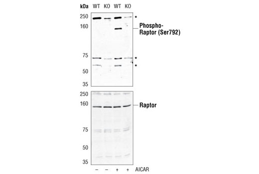
Western blot analysis of wild-type (WT) and AMPKα1 and α2 knockout (KO) mouse embryonic fibroblasts (MEFs), untreated or treated with AICAR (2 mM for 1 hour), using Phospho-Raptor (Ser792) Antibody (upper) or Raptor Antibody #4978 (lower). (Image provided by Dr. Reuben Shaw, Salk Institute for Biological Studies).
*Cross-reacting bands at 60, 70 and 240 kDa
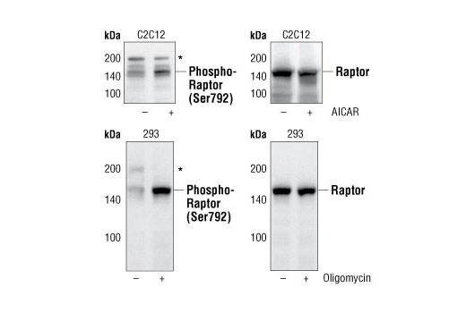
Western blot analysis of C2C12 or 293 cells, untreated or treated with AICAR (0.5 mM for 30 minutes) or oligomycin (0.5 μM for 30 minutes), using Phospho-Raptor (Ser792) Antibody (upper and lower left ) or Raptor Antibody #2280 (upper and lower right).
*Cross-reacting bands at 200 kDa.
Western blot analysis of extracts from various cell types using PRAS40 (D23C7) XP® Rabbit mAb.
Immunohistochemical analysis of paraffin-embedded human breast carcinoma using PRAS40 (D23C7) XP® Rabbit mAb.
Immunohistochemical analysis of paraffin-embedded human breast carcinoma, control (left) or λ phosphatase-treated (right), using Phospho-PRAS40 (Thr246) (C77D7) Rabbit mAb.
Immunohistochemical analysis of paraffin-embedded metastatic SKOV-3 tumor in mouse lung using Phospho-PRAS40 (Thr246) (C77D7) Rabbit mAb.
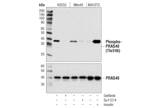
Western blot analysis of extracts from serum starved H3255, Mkn45 and NIH/3T3 cells, untreated or treated with either Iressa (1 μM, 3 hours), Su11274 (1 μM, 3 hours) or insulin (150 nM, 15 minutes), using Phospho-PRAS40 (Thr246) (C77D7) Rabbit mAb (upper) or PRAS40 (D23C7) Rabbit mAb #2691 (lower).
Western blot analysis of extracts from serum starved HeLa cells, untreated or treated with insulin (100 nM, 5 minutes) or with insulin and λ phosphatase, using Phospho-PRAS40 (Thr246) (C77D7) Rabbit mAb (upper) or PRAS40 Antibody #2610 (lower).
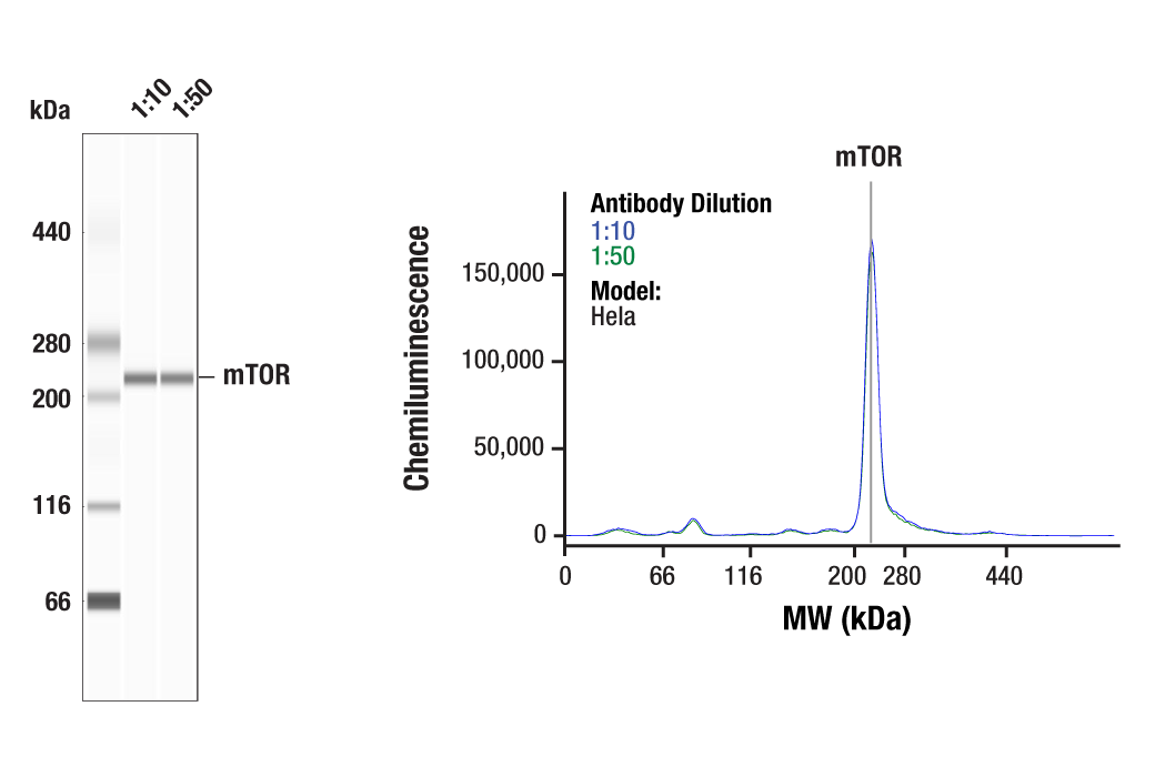
Western blot analysis of extracts from various cell types using mTOR (7C10) Rabbit mAb #2983.western blot 分析不同细胞系提取物,所用抗体为mTOR (7C10) Rabbit mAb #2983。
Western blot analysis of C2C12 or 293 cells, untreated or treated with AICAR (0.5 mM for 30 minutes) or oligomycin (0.5 μM for 30 minutes), using Phospho-Raptor (Ser792) Antibody #2083 (upper and lower left) or Raptor Antibody #2280 (upper and lower right). *Cross-reacting bands at 200 kDa.
Western blot分析C2C12 或293 cells,未处理组或AICAR (0.5 mM for 30 分钟) 或 oligomycin (0.5 μM for 30 分钟)处理组,所用抗体为Phospho-Raptor (Ser792) Antibody #2083 (上 和左下图) 或 Raptor Antibody #2280 (上和右下图). *与200 kDa的条带发生交叉反应。
Western blot analysis of extracts from serum starved H3255, Mkn45 and NIH/3T3 cells, untreated or treated with either Iressa (1 μM, 3 hours), Su11274 (1 μM, 3 hours) or insulin (150 nM, 15 minutes), using Phospho-PRAS40 (Thr246) (C77D7) Rabbit mAb #2997 (upper) or PRAS40 (D23C7) Rabbit mAb #2691 (lower).
western blot 分析血清饥饿的H3255, Mkn45 和NIH/3T3细胞提取物,未处理组和Iressa (1 μM, 3 小时), Su11274 (1 μM, 3 小时)或胰岛素 (150 nM, 15 分钟)处理组。所用抗体为 Phospho-PRAS40 (Thr246) (C77D7) Rabbit mAb #2997兔单抗 (上) 或 PRAS40 (D23C7) Rabbit mAb兔单抗 #2691 (下)
Western blot analysis of extracts from various cell types using PRAS40 (D23C7) XP® Rabbit mAb #2691.
western blot 分析不同细胞系提取物,所用抗体为PRAS40 (D23C7) XP® Rabbit mAb 兔单抗#2691.
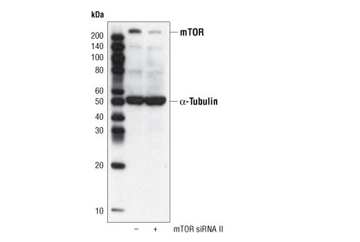
Western blot analysis of extracts from HeLa cells, transfected with 100 nM SignalSilence® Control siRNA (Fluorescein Conjugate) #6201 (-) or SignalSilence® mTOR siRNA II (+), using mTOR (7C10) Rabbit mAb #2983 and α-Tubulin (11H10) Rabbit mAb #2125. mTOR (7C10) Rabbit mAb confirms silencing of mTOR expression, while the α-Tubulin (11H10) Rabbit mAb is used to control for loading and specificity of mTOR siRNA.
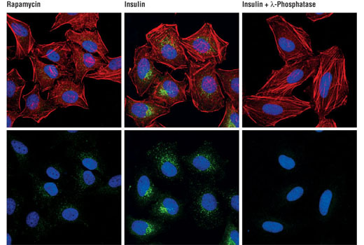
Confocal immunofluorescent analysis of HeLa cells, rapamycin-treated (#9904, 10 μM for 2 hours, left), insulin-treated (150 nM for 6 minutes, middle) or insulin- and λ-phosphatase-treated (right), using Phospho-mTOR (Ser2448) (D9C2) XP® Rabbit mAb (green). Actin filaments were labeled with DY-554 phalloidin. Blue pseudocolor = DRAQ5® #4084 (fluorescent DNA dye).
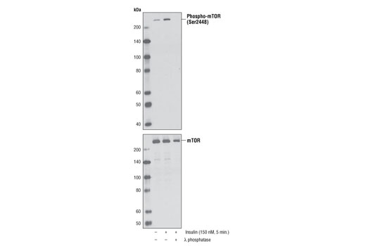
Western blot analysis of extracts from serum-starved NIH/3T3 cells, untreated or insulin-treated (150 nM, 5 minutes), alone or in combination with λ-phosphatase, using Phospho-mTOR (Ser2448) (D9C2) XP® Rabbit mAb (upper) or mTOR (7C10) Rabbit mAb #2983.
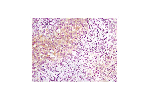
Immunohistochemical analysis of paraffin-embedded human lung carcinoma using RagC (D31G9) XP® Rabbit mAb.

 危险品化学品经营许可证(不带存储) 许可证编号:沪(杨)应急管危经许[2022]202944(QY)
  营业执照(三证合一)