


 全部商品分类
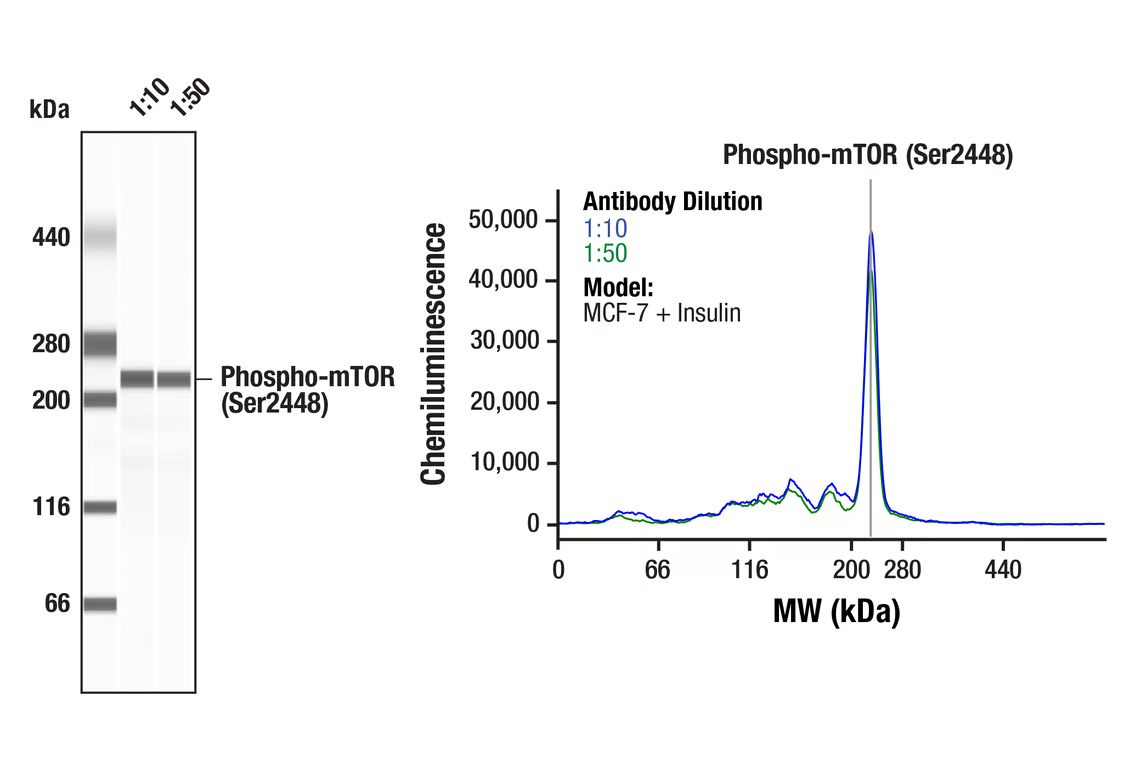


mTOR Pathway Antibody Sampler Kit

 下载产品说明书 下载SDS
 用小程序,查商品更便捷


 收藏
收藏
 对比
对比 咨询
咨询
The mTOR Pathway Antibody Sampler Kit contains reagents to investigate the control of protein translation, cell growth, and proliferation through mTOR signaling within cells. The kit contains enough primary and secondary antibodies to perform two Western blot experiments per primary antibody.
参考图片
Simple WesternTM analysis of lysates (0.1 mg/mL) from MCF-7 cells treated with insulin (100nM, 10 minutes) using Phospho-mTOR (Ser2448) (D9C2) XP® Rabbit mAb #5536. The virtual lane view (left) shows a single target band (as indicated) at 1:10 and 1:50 dilutions of primary antibody. The corresponding electropherogram view (right) plots chemiluminescence by molecular weight along the capillary at 1:10 (blue line) and 1:50 (green line) dilutions of primary antibody. This experiment was performed under reducing conditions on the JessTM Simple Western instrument from ProteinSimple, a BioTechne brand, using the 66-440 kDa separation module.
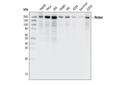
Western blot analysis of extracts from various cell lines, using Rictor (53A2) Rabbit mAb.
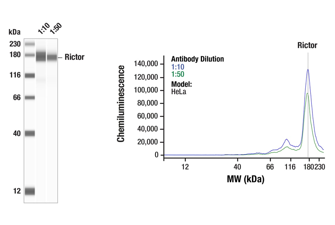
Simple WesternTM analysis of lysates (1 mg/mL) from HeLa cells using Rictor (53A2) Rabbit mAb #2114. The virtual lane view (left) shows a single target band (as indicated) at 1:10 and 1:50 dilutions of primary antibody. The corresponding electropherogram view (right) plots chemiluminescence by molecular weight along the capillary at 1:10 (blue line) and 1:50 (green line) dilutions of primary antibody. This experiment was performed under reducing conditions on the JessTM Simple Western instrument from ProteinSimple, a BioTechne brand, using the 12-230 kDa separation module.
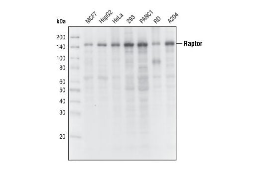
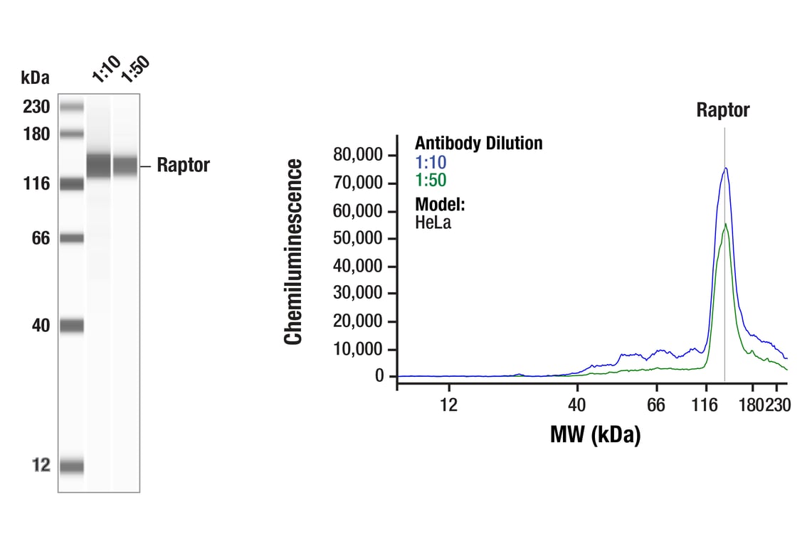
Western blot analysis of extracts from various cell lines, using Raptor (24C12) Rabbit mAb.
Simple WesternTM analysis of lysates (1 mg/ml) from HeLa cells using Raptor (24C12) Rabbit mAb #2280. The virtual lane view (left) shows the target band (as indicated) at 1:10 and 1:50 dilutions of primary antibody. The corresponding electropherogram view (right) plots chemiluminescence by molecular weight along the capillary at 1:10 (blue line) and 1:50 (green line) dilutions of primary antibody. This experiment was performed under reducing conditions on the JessTM Simple Western instrument from ProteinSimple, a BioTechne brand, using the 12-230kDa.
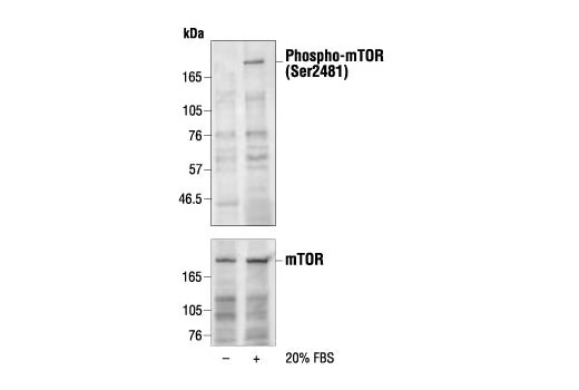
Western blot analysis of extracts from 293 cells (starved for 30 hours), untreated or treated with 20% FBS for 30 minutes, using Phospho-mTOR (Ser2481) Antibody (upper) or control mTOR Antibody #2972 (lower).
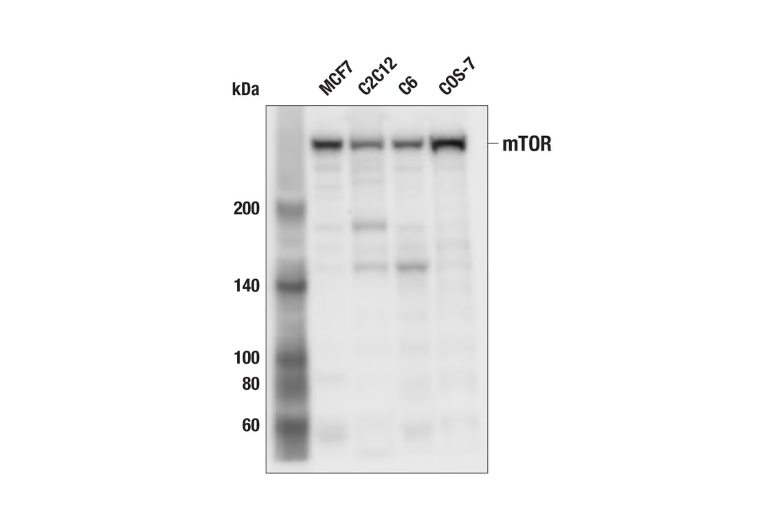
Western blot analysis of extracts from various cell lines using mTOR (7C10) Rabbit mAb.
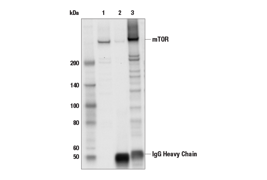
Immunoprecipitation of mTOR protein from MCF-7 cell extracts. Lane 1 is 10% input, lane 2 is Rabbit (DA1E) mAb IgG XP® Isotype Control #3900, and lane 3 is mTOR (7C10) Rabbit mAb. Western blot analysis was performed using mTOR (7C10) Rabbit mAb. Anti-rabbit IgG, HRP-linked Antibody #7074 was used as the secondary antibody.
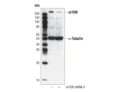
Western blot analysis of extracts from HeLa cells, transfected with 100 nM SignalSilence® Control siRNA (Fluorescein Conjugate) #6201 (-) or SignalSilence® mTOR siRNA II (+), using mTOR (7C10) Rabbit mAb #2983 and α-Tubulin (11H10) Rabbit mAb #2125. mTOR (7C10) Rabbit mAb confirms silencing of mTOR expression, while the α-Tubulin (11H10) Rabbit mAb is used to control for loading and specificity of mTOR siRNA.
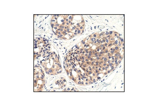
Immunohistochemical analysis of paraffin-embedded human breast carcinoma, showing cytoplasmic localization using mTOR (7C10) Rabbit mAb.
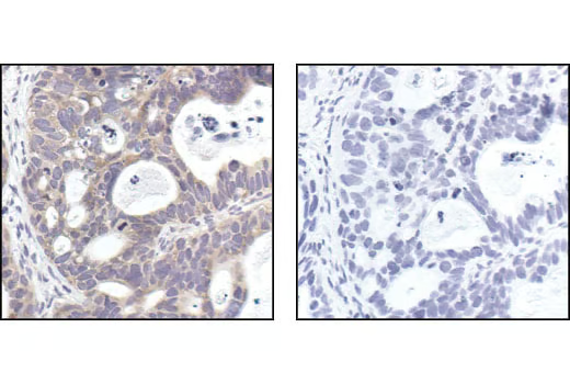
Immunohistochemical analysis of paraffin-embedded human lung carcinoma, using mTOR (7C10) Rabbit mAb in the presence of control peptide (left) or mTOR Blocking Peptide #1072 (right).
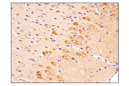
Immunohistochemical analysis of paraffin-embedded mouse brain using mTOR (7C10) Rabbit mAb.
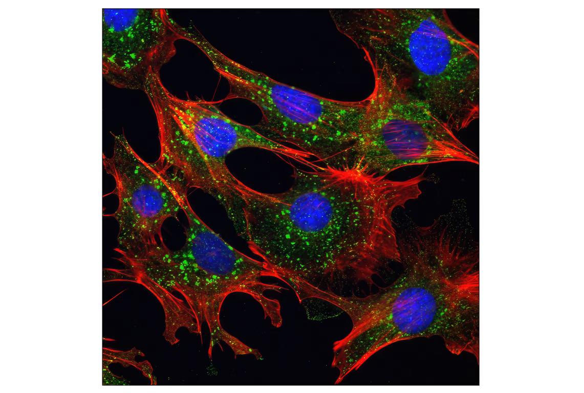
Confocal immunofluorescent analysis of mouse embryonic fibroblast (MEF) cells using mTOR (7C10) Rabbit mAb (green). Actin filaments were labeled with DY-554 phalloidin (red). Blue pseudocolor = DRAQ5® #4084 (fluorescent DNA dye).
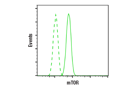
Flow cytometric analysis of A549 cells using mTOR (7C10) Rabbit mAb (solid line) compared to concentration-matched Rabbit (DA1E) mAb IgG XP® Isotype Control #3900 (dashed line). Anti-rabbit IgG (H+L), F(ab')2 Fragment (Alexa Fluor® 488 Conjugate) #4412 was used as a secondary antibody.
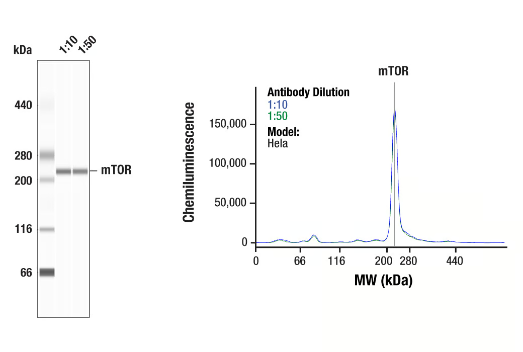
Simple Western™ analysis of lysates (0.1 mg/mL) from Hela cells using mTOR (7C10) Rabbit mAb #2983. The virtual lane view (left) shows a single target band (as indicated) at 1:10 and 1:50 dilutions of primary antibody. The corresponding electropherogram view (right) plots chemiluminescence by molecular weight along the capillary at 1:10 (blue line) and 1:50 (green line) dilutions of primary antibody. This experiment was performed under reducing conditions on the Jess™ Simple Western instrument from ProteinSimple, a BioTechne brand, using the 66-440 kDa separation module.
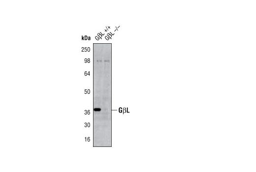
Western blot analysis of extracts from GβL wild-type (+/+) and knockout (-/-) mouse embryonic fibroblast cells using GβL (86B8) Rabbit mAb. (provided by the Whitehead Institute for Biomedical Research).
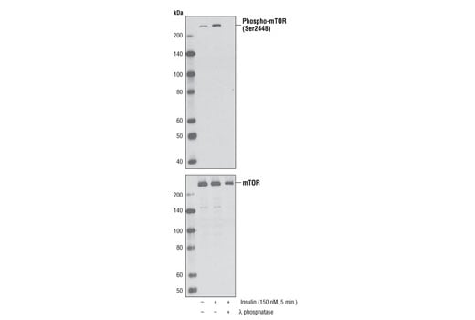
Western blot analysis of extracts from serum-starved NIH/3T3 cells, untreated or insulin-treated (150 nM, 5 minutes), alone or in combination with λ-phosphatase, using Phospho-mTOR (Ser2448) (D9C2) XP® Rabbit mAb (upper) or mTOR (7C10) Rabbit mAb #2983.
After the primary antibody is bound to the target protein, a complex with HRP-linked secondary antibody is formed. The LumiGLO® is added and emits light during enzyme catalyzed decomposition.
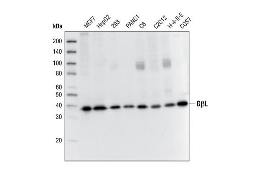
Western blot analysis of extracts from various cell lines, using GβL (86B8) Rabbit mAb.
Confocal immunofluorescent analysis of HeLa cells, rapamycin-treated (#9904, 10 nM for 2 hours, left), insulin-treated (150 nM for 6 minutes, middle) or insulin- and λ-phosphatase-treated (right), using Phospho-mTOR (Ser2448) (D9C2) XP® Rabbit mAb (green). Actin filaments were labeled with DY-554 phalloidin. Blue pseudocolor = DRAQ5® #4084 (fluorescent DNA dye).