


 全部商品分类
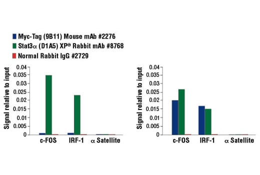


 用小程序,查商品更便捷


Monoclonal antibody is produced by immunizing animals with a synthetic peptide corresponding to residues 410-419 of human c-Myc (EQKLISEEDL).
Product Usage Information
For optimal ChIP results, use 5 μl of antibody and 10 μg of chromatin (approximately 4 x 10⁶ cells) per IP. This antibody has been validated using SimpleChIP® Enzymatic Chromatin IP Kits.
| Application | Dilution |
|---|---|
| Western Blotting | 1:1000 |
| Simple Western™ | 1:10 - 1:50 |
| Immunoprecipitation | 1:250 |
| Immunohistochemistry (Paraffin) | 1:1500 - 1:6000 |
| Immunofluorescence (Immunocytochemistry) | 1:1000 - 1:2000 |
| Flow Cytometry (Fixed/Permeabilized) | 1:500 - 1:2000 |
| Chromatin IP | 1:50 |
Specificity/Sensitivity
Species Reactivity:
All Species Expected
Supplied in 10 mM sodium HEPES (pH 7.5), 150 mM NaCl, 100 µg/ml BSA, 50% glycerol and less than 0.02% sodium azide. Store at –20°C. Do not aliquot the antibody.
For a carrier free (BSA and azide free) version of this product see product #58730.
参考图片
293T cells were either untransfected (left panel) or transfected with an Myc-tagged human Stat3 construct (right panel), then treated with Human Interleukin-6 (hIL-6) #8904 (100 ng/ml, 30 min). Chromatin immunoprecipitations were performed with cross-linked chromatin from cells and Myc-Tag (9B11) Mouse mAb #2276, Stat3α (D1A5) XP® Rabbit mAb #8768, or Normal Rabbit IgG #2729 using SimpleChIP® Enzymatic Chromatin IP Kit (Magnetic Beads) #9003. The enriched DNA was quantified by real-time PCR using SimpleChIP® Human c-Fos Promoter Primers #4663, human IRF-1 promoter primers, and SimpleChIP® Human α Satellite Repeat Primers #4486. The amount of immunoprecipitated DNA in each sample is represented as signal relative to the total amount of input chromatin, which is equivalent to one.
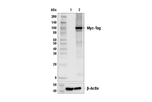
Western blot analysis of extracts from 293T cells, mock transfected (lane 1) or transiently transfected with a construct expressing Myc/DDK-tagged SARS-CoV-2 RNA-dependent RNA polymerase protein (lane 2) using Myc-Tag (9B11) Mouse mAb (upper) and β-Actin (D6A8) Rabbit mAb #8457 (lower).
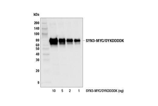
Western blot analysis of recombinant human SYN3-MYC/DYKDDDDK at various concentrations using, Myc-Tag (9B11) Mouse mAb.
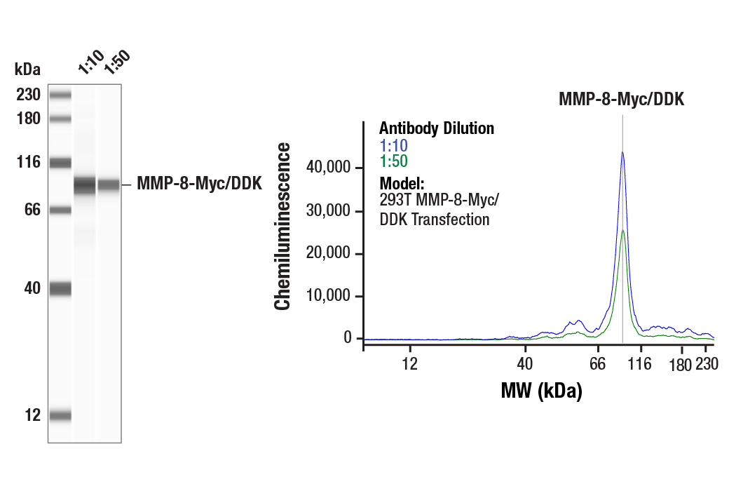
Simple Western™ analysis of lysates (0.1 mg/mL) from 293T cells, transfected with a construct expressing Myc/DDK tagged MMP-8 using Myc-Tag (9B11) Mouse mAb #2276. The virtual lane view (left) shows a single target band (as indicated) at 1:10 and 1:50 dilutions of primary antibody. The corresponding electropherogram view (right) plots chemiluminescence by molecular weight along the capillary at 1:10 (blue line) and 1:50 (green line) dilutions of primary antibody. This experiment was performed under reducing conditions on the Jess™ Simple Western instrument from ProteinSimple, a BioTechne brand, using the 12-230 kDa separation module.
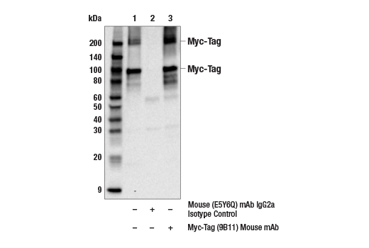
Immunoprecipitation of SARS-CoV-2 Spike-Myc/DDK protein from 293T transfected cell extracts. Lane 1 is 10% input, lane 2 is Mouse (E5Y6Q) mAb IgG2a Isotype Control #61656, and lane 3 is Myc-Tag (9B11) Mouse mAb. Western blot analysis was performed using Myc-Tag (71D10) Rabbit mAb #2278.
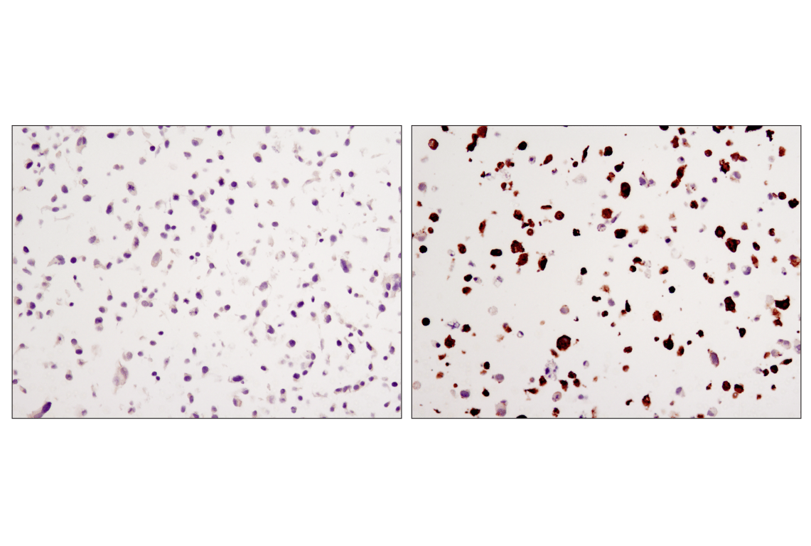
Immunohistochemical analysis of paraffin-embedded COS cell pellets, control (left) or transfected with a carboxy-terminal Myc tagged protein (right), using Myc-Tag (9B11) Mouse mAb.
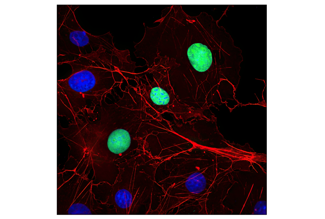
Confocal immunofluorescent analysis of COS cells transfected with a Myc-tagged protein using Myc-Tag (9B11) Mouse mAb (green). Actin filaments were labeled with DyLight™ 554 Phalloidin #13054 (red). Blue pseudocolor = DRAQ5® #4084 (fluorescent DNA dye).
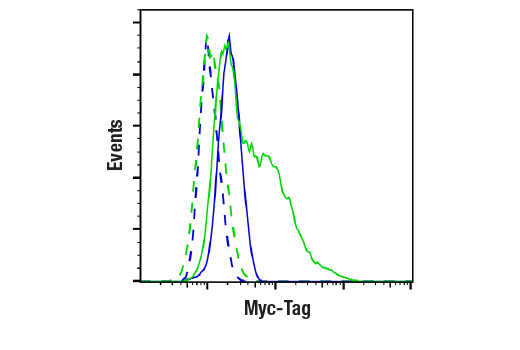
Flow cytometric analysis of 293T cells, mock-transfected (blue) or transfected with a construct expressing Myc-Tag (green), using Myc-Tag (9B11) Mouse mAb (solid lines) or concentration-matched Mouse (G3A1) mAb IgG1 Isotype Control #5415 (dashed lines). Anti-mouse IgG (H+L), F(ab')2 Fragment (Alexa Fluor® 488 Conjugate) #4408 was used as a secondary antibody.