The monoclonal antibody 29A1.4 specifically binds to mouse CD335, also known as NKp46. NKp46 is a 46 kDa type I transmembrane glycoprotein that is a member of the natural cytotoxicity receptor (NCR) family and immunoglobulin superfamily. NKp46 is encoded by the Ncr1 gene located on chromosome 7. NKp46 functions as a cytotoxicity triggering receptor and is selectively expressed by immature and mature NK cells in all mouse strains tested. NKp46 is detected on a minute fraction of NK-like T cells (less than 2% of NKp46+ express CD3e) but not on CD1d-restricted NKT cells from C57BL/6 mice. When immobilized on tissue culture plates, the 29A1.4 antibody reportedly stimulates NK cells to produce interferon-gamma and to release their cytoplasmic granule contents. Although the ligands for the NKp46 receptor have not been fully characterized, recent evidence indicates that this receptor plays an important role in the NK cell-mediated recognition and killing of some virus-infected cells and tumor cells. The immunogen used to generate the 29A1.4 clone was mouse NKp46-Fc recombinant protein.
商品描述
29A1.4
The monoclonal antibody 29A1.4 specifically binds to mouse CD335, also known as NKp46. NKp46 is a 46 kDa type I transmembrane glycoprotein that is a member of the natural cytotoxicity receptor (NCR) family and immunoglobulin superfamily. NKp46 is encoded by the Ncr1 gene located on chromosome 7. NKp46 functions as a cytotoxicity triggering receptor and is selectively expressed by immature and mature NK cells in all mouse strains tested. NKp46 is detected on a minute fraction of NK-like T cells (less than 2% of NKp46+ express CD3e) but not on CD1d-restricted NKT cells from C57BL/6 mice. When immobilized on tissue culture plates, the 29A1.4 antibody reportedly stimulates NK cells to produce interferon-gamma and to release their cytoplasmic granule contents. Although the ligands for the NKp46 receptor have not been fully characterized, recent evidence indicates that this receptor plays an important role in the NK cell-mediated recognition and killing of some virus-infected cells and tumor cells. The immunogen used to generate the 29A1.4 clone was mouse NKp46-Fc recombinant protein.
同种型
Rat IgG2a, κ
克隆号
克隆 29A1.4 (RUO)
浓度
0.2 mg/ml
产品详情
PE
R-Phycoerythrin (PE), is part of the BD family of Phycobiliprotein dyes. This fluorochrome is a multimeric fluorescent phycobiliprotein with excitation maximum (Ex Max) of 496 nm and 566 nm and an emission maximum (Em Max) at 576 nm. PE is designed to be excited by the Blue (488 nm), Green (532 nm) and Yellow-Green (561 nm) lasers and detected using an optical filter centered near 575 nm (e.g., a 575/26-nm bandpass filter). As PE is excited by multiple lasers, this can result in cross-laser excitation and fluorescence spillover on instruments with various combinations of Blue, Green, and Yellow-Green lasers. Please ensure that your instrument’s configurations (lasers and optical filters) are appropriate for this dye.
研发参考(4)
1. Biassoni R, Pessino A, Bottino C, Pende D, Moretta L, Moretta A. The murine homologue of the human NKp46, a triggering receptor involved in the induction of natural cytotoxicity. Eur J Immunol. 1999; 29(3):1014-1020. (Biology).
2. Gazit R, Gruda R, Elboim M, et al. Lethal influenza infection in the absence of the natural killer cell receptor gene Ncr1. Nat Immunol. 2006; 7(5):517-523. (Biology).
3. Joncker NT, Fernandez NC, Treiner E, Vivier E, Raulet DH. NK cell responsiveness is tuned commensurate with the number of inhibitory receptors for self-MHC class I: the rheostat model. J Immunol. 2009; 182(8):4572-4580. (Clone-specific: Flow cytometry).
4. Walzer T, Blery M, Chaix J, et al. Identification, activation, and selective in vivo ablation of mouse NK cells via NKp46. Proc Natl Acad Sci U S A. 2007; 104(9):3384-3389. (Clone-specific: Activation, Flow cytometry).
数据库链接
Entrez-Gene ID
17086
参考图片
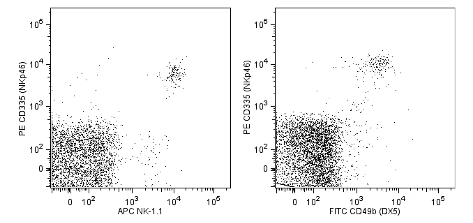
Flow cytometric analysis of PE anti-mouse CD335 (NKp46) expression on mouse splenocytes. C57BL/6 and BALB/c mouse spleen cells were stained separately with PE anti-mouse CD335 (NKp46) antibody. After washing, C57BL/6 cells were stained with APC-conjugated anti-mouse NK-1.1 (NKR-P1B and NKR-P1C) antibody (Cat. No.550627; left panel) and BALB/c cells were stained with FITC-conjugated anti-mouse CD49b (DX5) antibody (Cat. No.553857; right panel). Two-color dot plots showing the correlated expression patterns of CD335/NKp46 and either NK-1.1/CD161 (C57BL/6 cells; left panel) or DX5/CD49b (BALB/c cells; right panel) were derived from gated events with the forward and side light-scatter characteristics of viable lymphocytes. Flow cytometry was performed using a BD™ LSRII System.


 全部商品分类


 用小程序,查商品更便捷


