


 全部商品分类
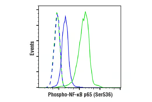


Phospho-NF-kappaB p65 (Ser536) (93H1) Rabbit mAb


 下载产品说明书 下载COA 下载SDS
 用小程序,查商品更便捷


 收藏
收藏
 对比
对比 咨询
咨询
Monoclonal antibody is produced by immunizing animals with a synthetic phosphopeptide corresponding to residues surrounding Ser536 of human NF-κB p65.
Product Usage Information
| Application | Dilution |
|---|---|
| Western Blotting | 1:1000 |
| Fluorescent Western | 1:1000 |
| Simple Western™ | 1:10 - 1:50 |
| Immunoprecipitation | 1:50 |
| Immunofluorescence (Immunocytochemistry) | 1:800 - 1:3200 |
| Flow Cytometry (Fixed/Permeabilized) | 1:800 - 1:3200 |
Specificity/Sensitivity
Species Reactivity:
Human, Mouse, Rat, Hamster, Monkey, Pig
Supplied in 10 mM sodium HEPES (pH 7.5), 150 mM NaCl, 100 µg/ml BSA, 50% glycerol and less than 0.02% sodium azide. Store at –20°C. Do not aliquot the antibody.
For a carrier free (BSA and azide free) version of this product see product #76778.
参考图片
Flow cytometric analysis of HeLa cells, untreated (blue) or treated with Human Tumor Necrosis Factor-α (hTNF-α) #8902 and Calyculin A #9902 (20 ng/ml and 100 nM, 15 min; green), using Phospho-NF-κB p65 (Ser536) (93H1) Rabbit mAb (solid lines) or concentration-matched Rabbit (DA1E) mAb IgG XP® Isotype Control #3900 (dashed lines). Anti-rabbit IgG (H+L), F(ab')2 Fragment (Alexa Fluor® 488 Conjugate) #4412 was used as a secondary antibody.
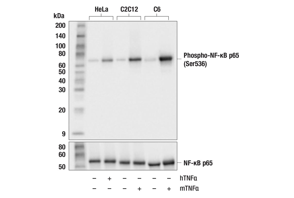
Western blot analysis of extracts from HeLa, C2C12, and C6 cells, untreated (-) or treated (+) as indicated with human TNF-α (hTNF⍺; 20ng/ml, 5min) or mouse TNF-α (mTNF⍺; 20ng/mL, 5min) using Phospho-NF-κB p65 (Ser536) (93H1) Rabbit mAb (upper) or NF-κB p65 (D14E12) XP® Rabbit mAb #8242 (lower). Phospho-NF-κB p65 is induced by human TNF-⍺ or mouse TNF-⍺ treatment as expected.
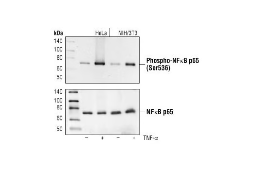
Western blot analysis of extracts from HeLa and NIH/3T3 cells, untreated or TNF-α treated (#2169, 20 ng/ml for 5 minutes), using Phospho-NF-κB p65 (Ser536) (93H1) Rabbit mAb (upper) or NF-κB p65 Antibody #3034 (lower).
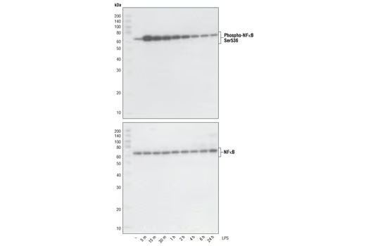
Western blot analysis of extracts from THP-1 cells, differentiated with TPA (#9905, 80 nM for 24h) and treated with 1 μg/ml LPS for the indicated times, using Phospho-NF-κB p65 (Ser536) (93H1) Rabbit mAb (upper) and NF-κB p65 (C22B4) Rabbit mAb #4764 (lower).
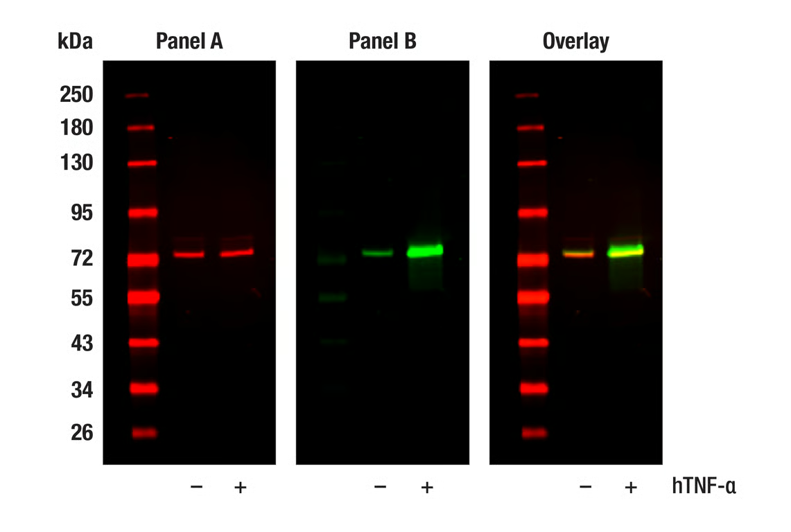
Western blot analysis of extracts from HeLa cells, untreated (-) or treated with hTNF-⍺ (20 ng/ml, 5 min; +), using NF-kB p65 (L8F6) Mouse mAb #6956 (Panel A) and Phospho-NF-kB p65 (Ser536) (93H1) Rabbit mAb #3033 (Panel B). Anti-mouse IgG (H+L) (DyLight 680 Conjugate) #5470 (red) and Anti-rabbit IgG (H+L) (DyLight 800 4X PEG Conjugate) #5151 (green) were used as secondary antibodies.
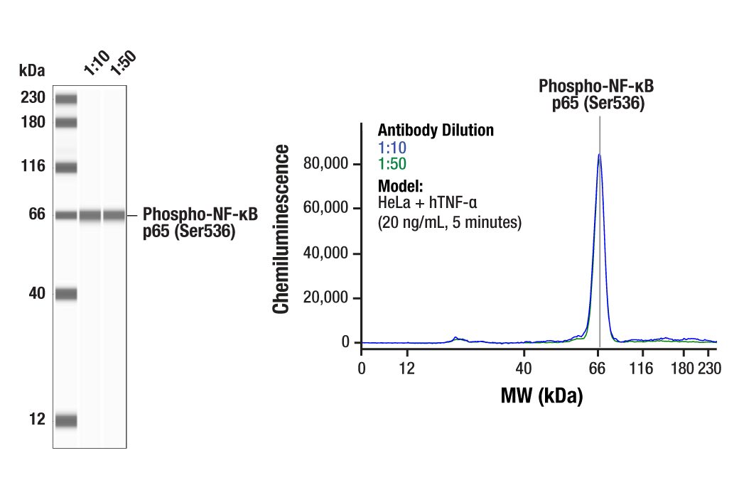
Simple Western™ analysis of lysates (1.0 mg/mL) from HeLa cells treated with hTNF-α (20 ng/mL, 5 minutes) using Phospho-NF-κB p65 (Ser536) (93H1) Rabbit mAb #3033. The virtual lane view (left) shows a single target band (as indicated) at 1:10 and 1:50 dilutions of primary antibody. The corresponding electropherogram view (right) plots chemiluminescence by molecular weight along the capillary at 1:10 (blue line) and 1:50 (green line) dilutions of primary antibody. This experiment was performed under reducing conditions on the Jess™ Simple Western instrument from ProteinSimple, a BioTechne brand, using the 12-230 kDa separation module.
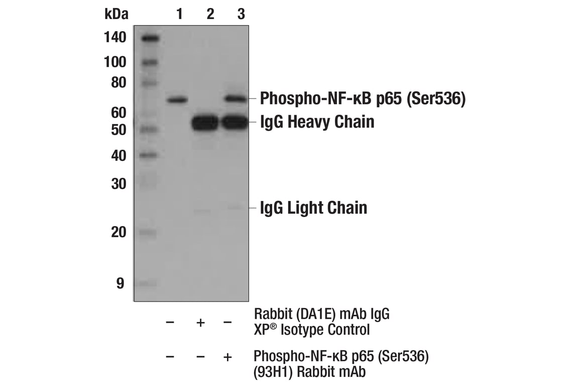
Immunoprecipitation of Phospho-NF-κB p65 (Ser536) from HeLa extracts treated with hTNF-α #8902 (20 ng/ml, 5 min). Lane 1 is 10% input, lane 2 is Rabbit (DA1E) mAb IgG XP® Isotype Control #3900, and lane 3 is Phospho-NF-κB p65 (Ser536) (93H1) Rabbit mAb. Western blot analysis was performed using Phospho-NF-κB p65 (Ser536) (93H1) Rabbit mAb. Anti-rabbit IgG, HRP-linked Antibody #7074 was used as a secondary antibody.
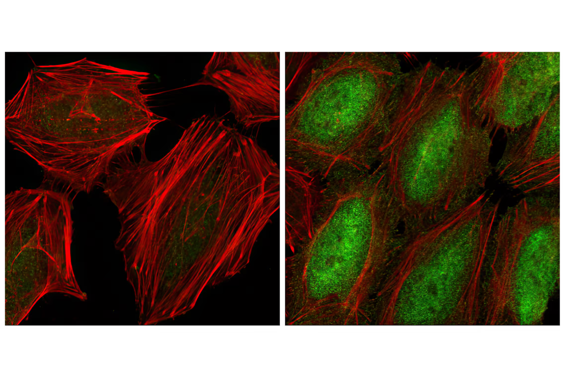
Confocal immunofluorescent analysis of HeLa cells, serum starved (left) or TNF-α treated (#8902 at 20 ng/ml for 20 min, right), using Phospho-NF-κB p65 (Ser536) (93H1) Rabbit mAb (green). Actin filaments have been labeled with Alexa Fluor® phalloidin 555 (red).