BD Phosflow™ BV421 Mouse Anti-Human NF-κB p65 (pS529)
1/3

BD Phosflow™ BV421 Mouse Anti-Human NF-κB p65 (pS529)

分享
品牌: BD Pharmingen
pdf 下载产品说明书
pdf 下载SDS
用小程序,查商品更便捷
收藏
对比对比
咨询咨询
    反应种属:
    Human (QC Testing)
    Human (QC Testing)
    来源宿主:
    Mouse BALB/c IgG2b, κ
    Mouse BALB/c IgG2b, κ
    展开
    产品介绍
    产品信息
    耦联标记
    BV421
    抗原名称
    NF-κB p65 (pS529)
    宿主
    Mouse BALB/c IgG2b, κ
    免疫原
    Phosphorylated Human NF-κB p65 Peptide
    简单描述
    The K10-895.12.50 monoclonal antibody recognizes the phosphorylated serine 529 (pS529) in the transactivation domain of the human NF- κ B p65 subunit. Nuclear factor κ B (NF- κ B) is a ubiquitously expressed transcription factor that regulates the expression of many other genes. It is crucial for cellular responses to a variety of stimuli including stress and microbial pathogens that lead to immunity, inflammation, proliferation, differentiation, survival, apoptosis, and tumorigenesis. The most studied NF- κ B complex consists of the p50 (also known as NF- κ B1) and p65 (also known as REL-A) subunits, both containing a 300-amino acid region with homology to the Rel proto-oncogene product (RH domain). The RH domain contains motifs for dimerization, nuclear localization, and binding to specific DNA sequences. In addition to the RH domain, the p65 subunit contains the transactivation domain, which is responsible for the interaction with the inhibitor I κ B and which contains phosphorylation sites. In most cell types, the p50/p65 heterodimer is located within the cytoplasm complexed to I κ B. This complex prevents nuclear translocation and activity of NF- κ B. In response to stimuli such as cytokines, LPS, DNA damage, and microbial infections, I κ B is phosphorylated at critical residues. This phosphorylation induces dissociation of the I κ B/NF- κ B complex, allowing the free heterodimeric NF- κ B to translocate to the nucleus. Furthermore, optimal activation of NF- κ B requires phosphorylation in the transactivation domain of p65. In the nucleus, activated NF- κ B dimers bind to the κ B sites within promoters and enhancers and function as transcriptional regulators. The antibody was conjugated to BD Horizon BV421 which is part of the BD Horizon Brilliant™ Violet family of dyes. With an Ex Max of 407-nm and Em Max at 421-nm, BD Horizon BV421 can be excited by the violet laser and detected in the standard Pacific Blue™ filter set (eg, 450/50-nm filter). BD Horizon BV421 conjugates are very bright, often exhibiting a 10 fold improvement in brightness compared to Pacific Blue conjugates.
    商品描述
    K10-895.12.50 The K10-895.12.50 monoclonal antibody recognizes the phosphorylated serine 529 (pS529) in the transactivation domain of the human NF- κ B p65 subunit. Nuclear factor κ B (NF- κ B) is a ubiquitously expressed transcription factor that regulates the expression of many other genes. It is crucial for cellular responses to a variety of stimuli including stress and microbial pathogens that lead to immunity, inflammation, proliferation, differentiation, survival, apoptosis, and tumorigenesis. The most studied NF- κ B complex consists of the p50 (also known as NF- κ B1) and p65 (also known as REL-A) subunits, both containing a 300-amino acid region with homology to the Rel proto-oncogene product (RH domain). The RH domain contains motifs for dimerization, nuclear localization, and binding to specific DNA sequences. In addition to the RH domain, the p65 subunit contains the transactivation domain, which is responsible for the interaction with the inhibitor I κ B and which contains phosphorylation sites. In most cell types, the p50/p65 heterodimer is located within the cytoplasm complexed to I κ B. This complex prevents nuclear translocation and activity of NF- κ B. In response to stimuli such as cytokines, LPS, DNA damage, and microbial infections, I κ B is phosphorylated at critical residues. This phosphorylation induces dissociation of the I κ B/NF- κ B complex, allowing the free heterodimeric NF- κ B to translocate to the nucleus. Furthermore, optimal activation of NF- κ B requires phosphorylation in the transactivation domain of p65. In the nucleus, activated NF- κ B dimers bind to the κ B sites within promoters and enhancers and function as transcriptional regulators. The antibody was conjugated to BD Horizon BV421 which is part of the BD Horizon Brilliant™ Violet family of dyes. With an Ex Max of 407-nm and Em Max at 421-nm, BD Horizon BV421 can be excited by the violet laser and detected in the standard Pacific Blue™ filter set (eg, 450/50-nm filter). BD Horizon BV421 conjugates are very bright, often exhibiting a 10 fold improvement in brightness compared to Pacific Blue conjugates.
    同种型
    Mouse BALB/c IgG2b, κ
    克隆号
    克隆 K10-895.12.50 (RUO)
    产品详情
    BV421
    The BD Horizon Brilliant Violet™ 421 (BV421) Dye is part of the BD Horizon Brilliant Violet™ family of dyes. This polymer-technology based dye has an excitation maximum (Ex Max) of 407-nm and an emission maximum (Em Max) at 423-nm. Driven by BD innovation, BV421 is designed to be excited by the violet laser (405-nm) and detected using an optical filter centered near 420-nm (e.g., a 431/28-nm or 450/50-nm bandpass filter). BV421 is an ideal alternative for V450 as it is approximately ten times brighter with less spillover into the BV510/V500 detector. Please ensure that your instrument’s configurations (lasers and optical filters) are appropriate for this dye.
    BV421
    Violet 405 nm
    407 nm
    423 nm
    应用
    实验应用
    Intracellular staining (flow cytometry) (Routinely Tested)
    推荐用量
    5 µl
    反应种属
    Human (QC Testing)
    目标/特异性
    NF-κB p65 (pS529)
    背景
    别名
    RELA; REL-A; NFKBp65; NFKB3; Transcription factor p65; TF65
    翻译后修饰
    Phosphate
    制备和贮存
    存储溶液
    Aqueous buffered solution containing BSA and ≤0.09% sodium azide.
    保存方式
    Aqueous buffered solution containing BSA and ≤0.09% sodium azide.
    文献
    文献
    研发参考(8) 1. Dominguez-Villar M, Gautron AS, de Marcken M, Keller MJ, Hafler DA. TLR7 induces anergy in human CD4+ T cells. Nat Immunol. 2015; 16(1):118-128. (Clone-specific: Flow cytometry). 2. Feasibility study: phospho-specific flow cytometry enabling rapid functional analysis of bone marrow samples from patients with multiple myeloma. Cytometry B Clin Cytom. 2014; 86B:139-144. (Clone-specific: Flow cytometry). 3. Mingueneau M, Kreslavsky T, Gray D, . The transcriptional landscape of alphabeta T cell differentiation. Nat Immunol. 2013; 14(6):619-632. (Clone-specific: Flow cytometry). 4. Natoli G, Saccani S, Bosisio D, Marazzi I. Interactions of NF-kappaB with chromatin: the art of being at the right place at the right time. Nat Immunol. 2005; 6(5):439-445. (Biology). 5. Siebenlist U, Brown K, Claudio E. Control of lymphocyte development by nuclear factor-kappaB. Nat Rev Immunol. 2005; 5:435-445. (Biology). 6. Suni MA, Maino VC. Flow cytometric analysis of cell signaling proteins. Methods Mol Biol. 2011; 717:155-169. (Clone-specific: Flow cytometry). 7. Viatour P, Merville M-P, Bours V, Chariot A. Phosphorylation of NF-kappaB and IkappaB proteins: implications in cancer and inflammation. Trends Biochem Sci. 2005; 30(1):43-52. (Biology). 8. van de Laar L, van den Bosch A, Boonstra A, et al. PI3K-PKB hyperactivation augments human plasmacytoid dendritic cell development and function. Blood. 2012; 120(25):4982-4991. (Clone-specific: Flow cytometry).

    参考图片

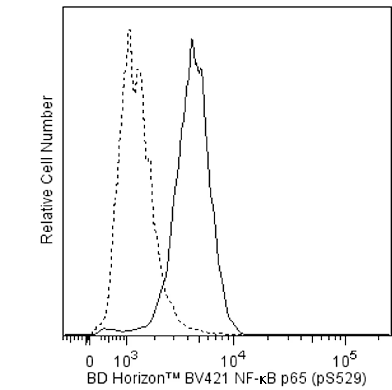
    Flow cytometric analysis of NF-κB p65 (pS529) expression by TNF-treated HeLa S3 cells. Cultured cells from the human HeLa S3 (Cervical adenocarcinoma, ATCC CCL 2.2) cell line were starved overnight in Dulbecco's Minimal Eagle's Medium. The cells were harvested and washed with Dulbecco's Phosphate Buffered Saline. They were then either left untreated (dashed line histogram) or treated (37°C, 10 min) with Recombinant Human TNF protein (20 ng/mL; Cat. No. 554618; solid line histogram) and Calyculin A (50 nM; Calbiochem Cat. No. 208851). The cells were fixed (10 min; 37°C) with pre-warmed BD Cytofix™ Fixation Buffer (Cat. No. 554655), permeabilized (30 min on ice) with BD Phosflow™ Perm Buffer III (Cat. No. 558050), and washed twice with BD Pharmingen™ Stain Buffer (FBS) (Cat. No. 554656). The cells were then stained with BD Phosflow™ BV421 Mouse Anti-Human NF-κB p65 (pS529) antibody (Cat. No. 565446).  The fluorescence histograms showing NF-κB p65 (pS529) expression were derived from gated events with the forward and side light-scatter characteristics of intact HeLa S3 cells. Flow cytometric analysis was performed using a BD™ LSR II Flow Cytometer System.

    声明 :本官网所有报价均为常温或者蓝冰运输价格,如有产品需要干冰运输,需另外加收干冰运输费。
    货号:
    565446
    一键复制
    询价
    供应商现货
    50Tst
    选择数量
    当前规格1件起购 
    配送至
    预计5-6个工作日送达,快递: 免运费,若需干冰额外收费