


 全部商品分类
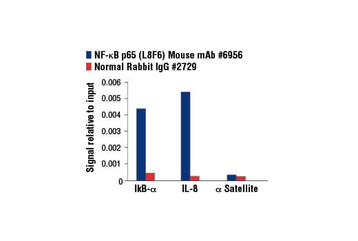


NF-kappaB p65 (L8F6) Mouse mAb

 下载产品说明书 下载COA 下载SDS
 用小程序,查商品更便捷


 收藏
收藏
 对比
对比 咨询
咨询
Monoclonal antibody is produced by immunizing animals with a synthetic peptide corresponding to residues near the carboxy terminus of human NF-κB protein.
Product Usage Information
For optimal ChIP results, use 10 μl of antibody and 10 μg of chromatin (approximately 4 x 10⁶ cells) per IP.This antibody has been validated using SimpleChIP® Enzymatic Chromatin IP Kits.
| Application | Dilution |
|---|---|
| Western Blotting | 1:1000 |
| Fluorescent Western | 1:1000 |
| Immunoprecipitation | 1:100 |
| Immunofluorescence (Immunocytochemistry) | 1:400 - 1:1600 |
| Flow Cytometry (Fixed/Permeabilized) | 1:200 - 1:800 |
| Chromatin IP | 1:50 |
Specificity/Sensitivity
Species Reactivity:
Human, Mouse, Rat, Hamster, Monkey, Mink, Bovine, Dog, Pig
Supplied in 10 mM sodium HEPES (pH 7.5), 150 mM NaCl, 100 µg/ml BSA, 50% glycerol and less than 0.02% sodium azide. Store at –20°C. Do not aliquot the antibody.
For a carrier free (BSA and azide free) version of this product see product #64921.
参考图片
Chromatin immunoprecipitations were performed with cross-linked chromatin from HeLa cells treated with Human Tumor Necrosis Factor-α (hTNF-α) #8902 (30 ng/ml, 1 hr) and either NF-κB p65 (L8F6) Mouse mAb or Normal Rabbit IgG #2729 using SimpleChIP® Enzymatic Chromatin IP Kit (Magnetic Beads) #9003. The enriched DNA was quantified by real-time PCR using SimpleChIP® Human IκBα Promoter Primers #5552, human IL-8 promoter primers, and SimpleChIP® Human α Satellite Repeat Primers #4486. The amount of immunoprecipitated DNA in each sample is represented as signal relative to the total amount of input chromatin, which is equivalent to one.
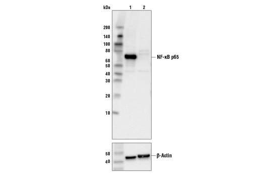
Western blot analysis of extracts from control HeLa cells (lane 1) or NF-κB p65 knockout HeLa cells (lane 2) using NF-κB p65 (L8F6) Mouse mAb #6956 (upper) or β-actin (13E5) Rabbit mAb #4970 (lower). The absence of signal in the NF-κB p65 knockout HeLa cells confirms the specificity of the antibody for NF-κB p65.
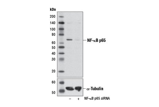
Western blot analysis of extracts from HeLa cells, transfected with 100 nM SignalSilence® Control siRNA (Unconjugated) #6568 (-) or SignalSilence® NF-κB p65 siRNA I #6261 (+), using NF-κB p65 (L8F6) Mouse mAb (upper) or α-Tubulin (11Η10) Rabbit mAb #2125 (lower). The NF-κB p65 (L8F6) Mouse mAb confirms silencing of NF-κB p65 expression, while the α-Tubulin (11Η10) Rabbit mAb is used as a loading control.
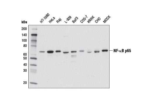
Western blot analysis of extracts from various cell lines using NF-κB p65 (L8F6) Mouse mAb.
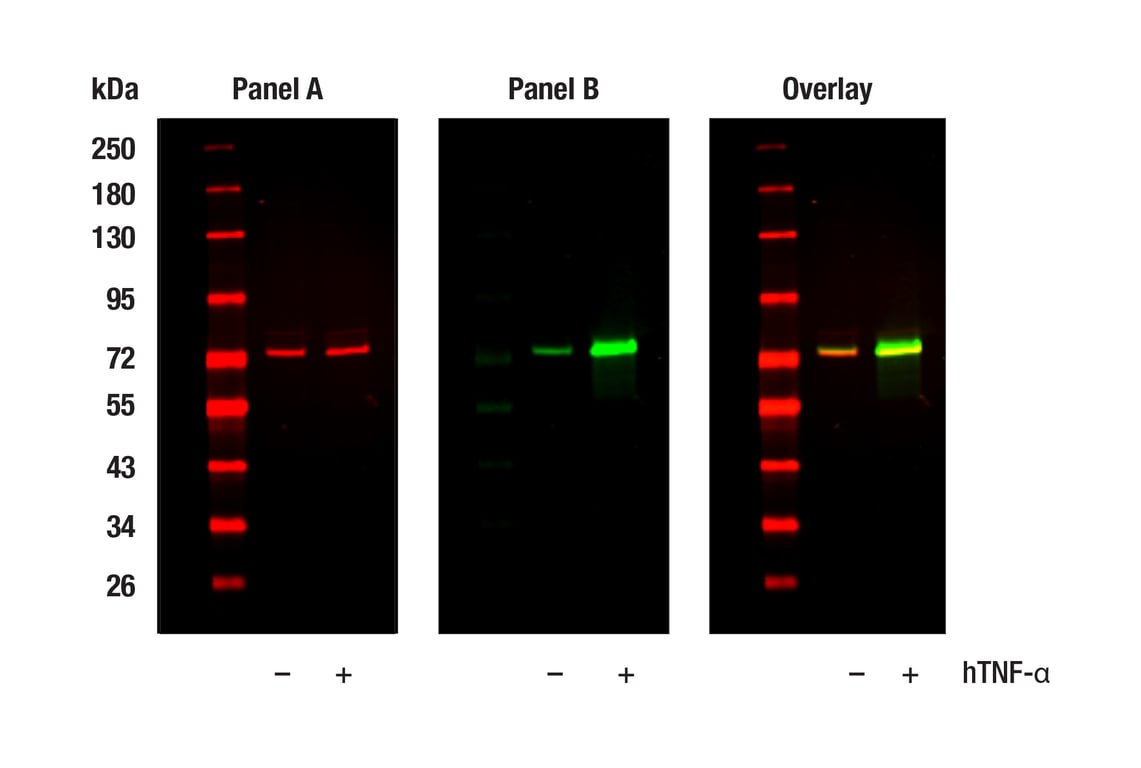
Western blot analysis of extracts from HeLa cells, untreated (-) or treated with hTNF-⍺ (20 ng/ml, 5 min; +), using NF-kB p65 (L8F6) Mouse mAb #6956 (Panel A) and Phospho-NF-kB p65 (Ser536) (93H1) Rabbit mAb #3033 (Panel B). Anti-mouse IgG (H+L) (DyLight 680 Conjugate) #5470 (red) and Anti-rabbit IgG (H+L) (DyLight 800 4X PEG Conjugate) #5151 (green) were used as secondary antibodies.
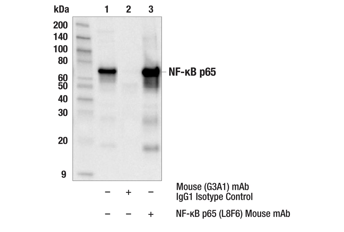
Immunoprecipitation of NF-kB p65 from HeLa cell extracts. Lane 1 is 10% input, lane 2 is precipitated with Mouse (G3A1) mAb IgG1 Isotype Control #5415, and lane 3 is NF-κB p65 (L8F6) Mouse mAb, #6956. Western blot was performed using NF-κB p65 (D14E12) XP® Rabbit mAb, #8242.
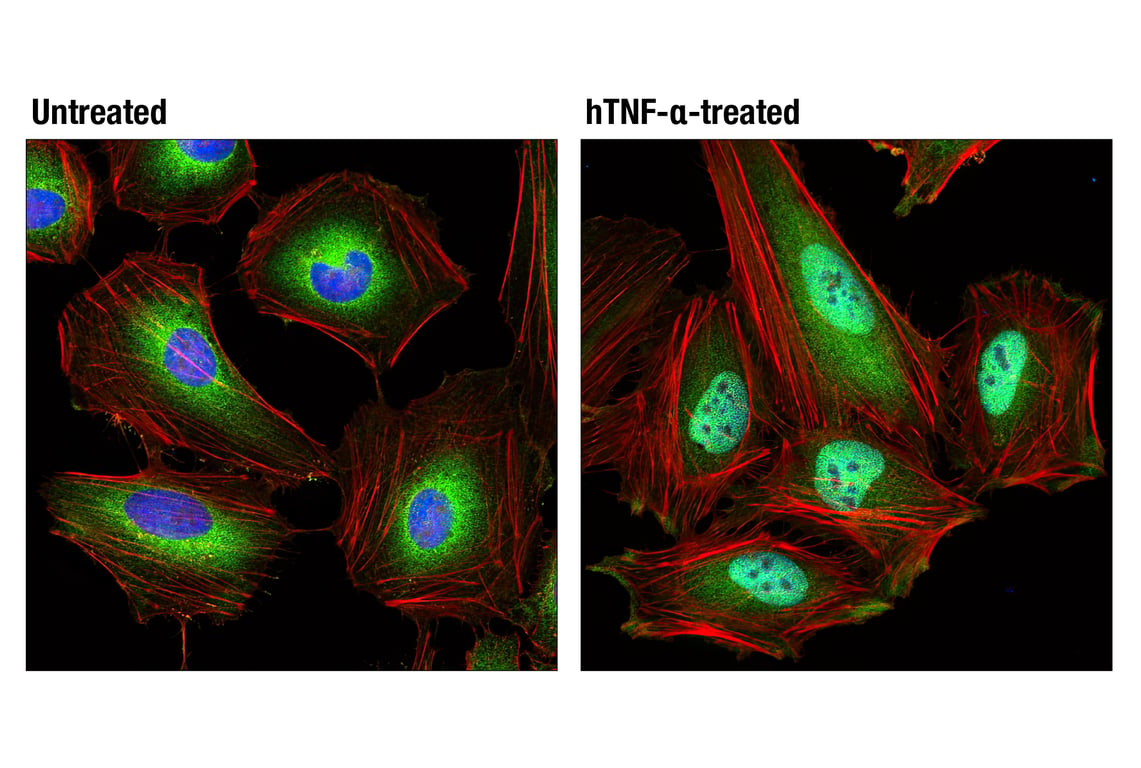
Confocal immunofluorescent analysis of HeLa cells, untreated (left) or treated with Human Tumor Necrosis Factor-α (hTNF-α) #8902 (20 ng/mL, 20 min; right), using NF-κB p65 (L8F6) Mouse mAb (green). Actin filaments were labeled with DY-554 phalloidin (red). Blue pseudocolor = DRAQ5® #4084 (fluorescent DNA dye).
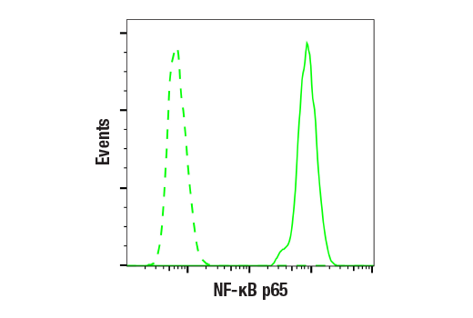
Flow cytometric analysis of MCF7 cells using NF-kB p65 (L8F6) Mouse mAb (solid line) compared to a concentration-matched Mouse (G3A1) mAb IgG1 Isotype Control #5415 (dashed lines). Anti-mouse IgG (H+L), F(ab')2 Fragment (Alexa Fluor® 488 Conjugate) #4408 was used as a secondary antibody.