


 全部商品分类
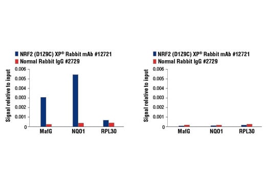


 用小程序,查商品更便捷


Monoclonal antibody is produced by immunizing animals with a synthetic peptide corresponding to residues surrounding Ala275 of human NRF2 protein.
Product Usage Information
For optimal ChIP and ChIP-seq results, use 5 μl of antibody and 10 μg of chromatin (approximately 4 x 10⁶ cells) per IP. This antibody has been validated using SimpleChIP® Enzymatic Chromatin IP Kits.
| Application | Dilution |
|---|---|
| Western Blotting | 1:1000 |
| Immunoprecipitation | 1:50 |
| Immunofluorescence (Immunocytochemistry) | 1:200 - 1:800 |
| Flow Cytometry (Fixed/Permeabilized) | 1:1600 - 1:6400 |
| Chromatin IP | 1:200 |
| Chromatin IP-seq | 1:200 |
Specificity/Sensitivity
Species Reactivity:
Human, Mouse, Monkey
Supplied in 10 mM sodium HEPES (pH 7.5), 150 mM NaCl, 100 µg/ml BSA, 50% glycerol and less than 0.02% sodium azide. Store at –20°C. Do not aliquot the antibody.
For a carrier free (BSA and azide free) version of this product see product #84599.
参考图片
Chromatin immunoprecipitations were performed with cross-linked chromatin from MEF NRF2 wild-type (left) and NRF2 knock-out (right) cells, both treated with DEM (50 μM, 3 hr), and NRF2 (D1Z9C) XP® Rabbit mAb or Normal Rabbit IgG #2729 using SimpleChIP® Enzymatic Chromatin IP Kit (Magnetic Beads) #9003. The enriched DNA was quantified by real-time PCR using mouse MafG intron 1 primers, SimpleChIP® Mouse NQO1 Promoter Primers #12635, and SimpleChIP® Mouse RPL30 Intron 2 Primers #7015. The amount of immunoprecipitated DNA in each sample is represented as signal relative to the total amount of input chromatin, which is equivalent to one.
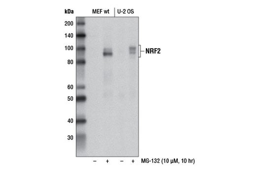
Western blot analysis of extracts from MEF wt and U-2 OS cells, untreated (-) or treated with MG-132 #2194 (10 μM, 10 hr; +), using NRF2 (D1Z9C) XP® Rabbit mAb.
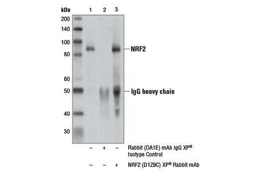
Immunoprecipitation of NRF2 from MEF wt cell extracts treated with MG-132 #2194 (10 μM, 10 hr) using Rabbit (DA1E) mAb IgG XP® Isotype Control #3900 (lane 2) or NRF2 (D1Z9C) XP® Rabbit mAb (lane 3). Lane 1 is 10% input. Western blot analysis was performed using NRF2 (D1Z9C) XP® Rabbit mAb (lane 3).
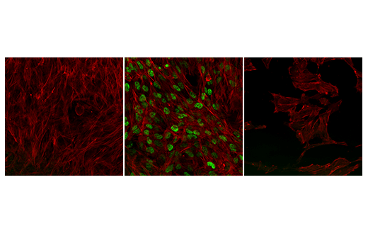
Confocal immunofluorescent analysis of wild-type NRF2 MEF cells, untreated (left) or treated with MG-132 #2194 (10 μM, 8 hr; center), and NRF2 knock-out MEF cells treated with MG-132 #2194 (10 μM, 8 hr; right), using NRF2 (D1Z9C) XP® Rabbit mAb (green pseudocolor). Actin filaments were labeled with Alexa Fluor® 488 Phalloidin #8878 (red pseudocolor).
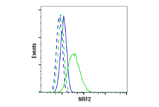
Flow cytometric analysis of MEF wt cells, untreated (blue) or treated with MG-132 #2194 (10uM, 4 hrs; green) using NRF2 (D1Z9C) XP® Rabbit mAb (solid line) compared to concentration-matched Rabbit (DA1E) mAb IgG XP® Isotype Control #3900 (dashed line). Anti-rabbit IgG (H+L), F(ab')2 Fragment (Alexa Fluor® 488 Conjugate) #4412 was used as a secondary antibody.
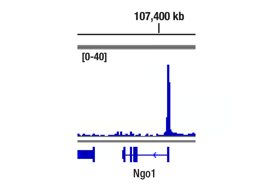
Chromatin immunoprecipitations were performed with cross-linked chromatin from MEF cells treated with DEM (50 μM, 3 hr) and NRF2 (D1Z9C) XP® Rabbit mAb, using SimpleChIP® Plus Enzymatic Chromatin IP Kit (Magnetic Beads) #9005. DNA Libraries were prepared using DNA Library Prep Kit for Illumina Systems (ChIP-seq, CUT&RUN) #56795. The figure shows binding across NQO1, a known target gene of NRF2 (see additional figure containing ChIP-qPCR data).
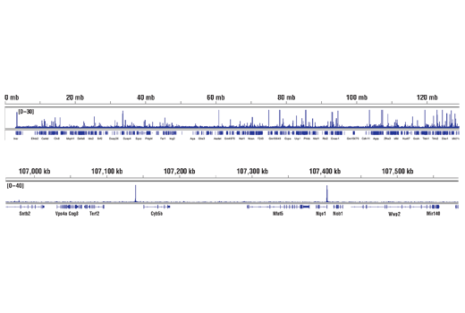
Chromatin immunoprecipitations were performed with cross-linked chromatin from MEF cells treated with DEM (50 μM, 3 hr) and NRF2 (D1Z9C) XP® Rabbit mAb, using SimpleChIP® Plus Enzymatic Chromatin IP Kit (Magnetic Beads) #9005. DNA Libraries were prepared using DNA Library Prep Kit for Illumina® (ChIP-seq, CUT&RUN) #56795. The figure shows binding across chromosome 8 (upper), including NQO1 (lower), a known target gene of NRF2 (see additional figure containing ChIP-qPCR data).