


 全部商品分类
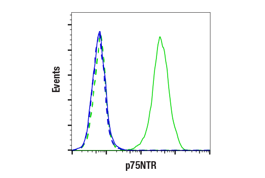


p75NTR (D4B3) XP ® Rabbit mAb

 下载产品说明书 下载COA 下载SDS
 用小程序,查商品更便捷


 收藏
收藏
 对比
对比 咨询
咨询
Monoclonal antibody is produced by immunizing animals with a synthetic peptide corresponding to residues surrounding Arg198 of human p75NTR protein. This antibody is predicted to bind the extracellular amino-terminal region of p75NTR protein.
Product Usage Information
| Application | Dilution |
|---|---|
| Western Blotting | 1:1000 |
| Immunoprecipitation | 1:50 |
| Immunofluorescence (Frozen) | 1:1600 - 1:3200 |
| Immunofluorescence (Immunocytochemistry) | 1:1600 - 1:3200 |
| Flow Cytometry (Live) | 1:200 - 1:800 |
Specificity/Sensitivity
Species Reactivity:
Human, Mouse, Rat
Supplied in 10 mM sodium HEPES (pH 7.5), 150 mM NaCl, 100 µg/ml BSA, 50% glycerol and less than 0.02% sodium azide. Store at –20°C. Do not aliquot the antibody.
For a carrier free (BSA and azide free) version of this product see product #46333.
参考图片
Flow cytometric analysis of live DLD-1 cells (blue, negative) and SK-N-MC cells (green, positive) using p75NTR (D4B3) Rabbit mAb (solid lines) or a concentration-matched Rabbit (DA1E) mAb IgG XP® Isotype Control #3900 (dashed lines). Anti-rabbit IgG (H+L), F(ab')2 Fragment (Alexa Fluor® 488 Conjugate) #4412 was used as a secondary antibody.
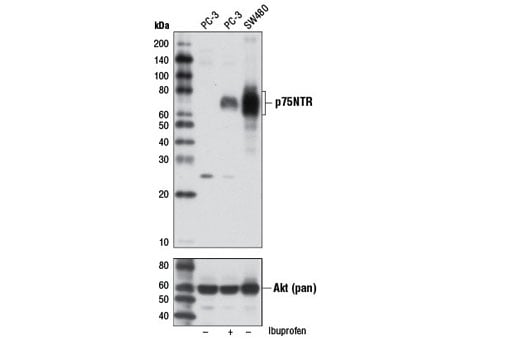
Western blot analysis of extracts from PC-3 cells, untreated (-) or ibuprofen-treated (2 mM, 24 hr) (+), and SW480 cells (-) using p75NTR (D4B3) XP® Rabbit mAb (upper) and Akt (pan) (C67E7) Rabbit mAb #4691 (lower).
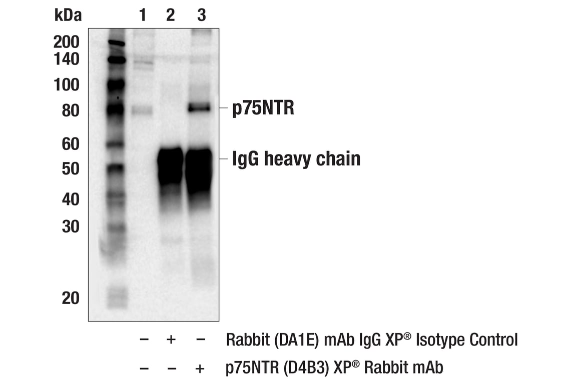
Immunoprecipitation of p75NTR from SW-480 cell extracts. Lane 1 is 10% input, lane 2 is Rabbit (DA1E) mAb IgG XP® Isotype Control #3900, and lane 3 is p75NTR (D4B3) XP® Rabbit mAb. Western blot was performed using p75NTR (D4B3) XP® Rabbit mAb.
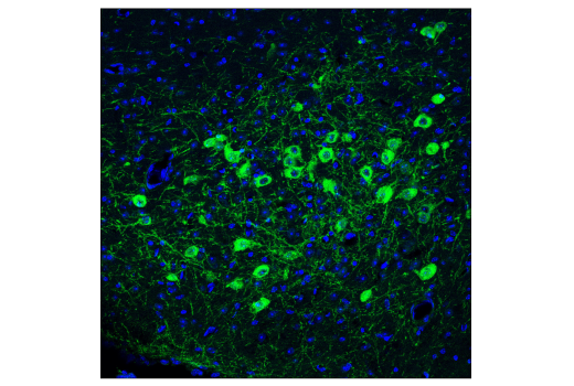
Confocal immunofluorescent analysis of fixed frozen mouse optic tract labeled with p75NTR (D4B3) XP® Rabbit mAb (green) and DAPI #8961 (blue).
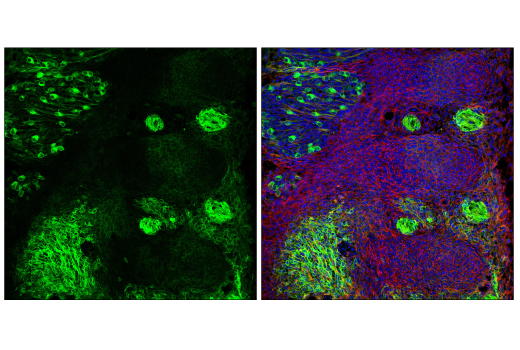
Confocal immunofluorescent analysis of fixed frozen developing mouse E12 spinal cord and somites labeled with p75NTR (D4B3) XP® Rabbit mAb (left, green), DyLight™ 554 Phalloidin #13054 (right, red), and DAPI #8961 (right, blue).
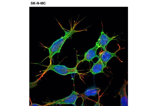
Confocal immunofluorescent analysis of SK-N-MC cells using p75NTR (D4B3) XP® Rabbit mAb (green). Actin filaments were labeled with DY-554 phalloidin (red). Blue pseudocolor = DRAQ5® #4084 (fluorescent DNA dye).