


 全部商品分类


1/2
BD Pharmingen™ PE-Cy™7 Mouse Anti-Mouse NK-1.1

品牌: BD Pharmingen
 下载产品说明书
 用小程序,查商品更便捷


 收藏
收藏
 对比
对比 咨询
咨询反应种属:
Mouse (QC Testing)
来源宿主:
Mouse C3H x BALB/c IgG2a, κ
产品介绍
产品使用步骤推荐 产品介绍
产品信息
荧光素标记
抗原名称
NK1.1

宿主
Mouse C3H x BALB/c IgG2a, κ
免疫原
Mouse NK-1+ Spleen and Bone Marrow Cells
简单描述
In the mouse, at least three members of the
Klrb
(
K
iller cell
l
ectin-like
r
eceptor, subfamily
b
; formerly
NKR-P1
) gene family have been identified (
Klrb1a/NKR-P1A, Klrb1b/NKR-P1B,
and
Klrb1c/NKR-P1C
); but in the human gene family, a single homologue has been designated
KLRB1, NKR-P1A, or CD161
. The KLRB1/NKR-P1 family of proteins are type-II-transmembrane C-type lectin receptors. KLRB1C/NKR-P1C activates NK-cell cytotoxicity, while KLRB1B/NKR-P1B functions as an inhibitory receptor. KLRB1B/NKR-P1B protein has intracellular
I
mmunoreceptor
T
yrosine-based
I
nhibitory
M
otif (ITIM), while KLRB1C/NKR-P1C lacks ITIM and activates via association with Fc Receptor γ chain. Strikingly, KLRB1B/NKR-P1B and KLRB1C/NKR-P1C share 96% amino acid sequence identity in their extracellular C-type lectin domains. The PK136 antibody reacts with the NK-1.1 surface antigen (CD161c) encoded by the
Klrb1c/NKR-P1C
gene expressed on natural killer (NK) cells in selected strains of mice (eg, C57BL, FVB/N, NZB, but not A, AKR, BALB/c, CBA/J, C3H, C57BR, C58, DBA/1, DBA/2, NOD, SJL, 129) and the CD161b antigen encoded by the
Klrb1b/NKR-P1B
gene expressed only on Swiss NIH and SJL mice, but not on C57BL/6. Expression of KLRB1C/NKR-P1C protein is correlated with the ability to lyse tumor cells in vitro and to mediate rejection of bone marrow allografts. The NK-1.1 marker is useful in defining NK cells; however, the antigen is also expressed on a rare, specialized population of T lymphocytes (NK-T cells) and some cultured monocytes. Plate-bound PK136 mAb, in combination with low concentrations of IL-2, induces proliferation of a subset of NK cells.
商品描述
PK136
In the mouse, at least three members of the
Klrb
(
K
iller cell
l
ectin-like
r
eceptor, subfamily
b
; formerly
NKR-P1
) gene family have been identified (
Klrb1a/NKR-P1A, Klrb1b/NKR-P1B,
and
Klrb1c/NKR-P1C
); but in the human gene family, a single homologue has been designated
KLRB1, NKR-P1A, or CD161
. The KLRB1/NKR-P1 family of proteins are type-II-transmembrane C-type lectin receptors. KLRB1C/NKR-P1C activates NK-cell cytotoxicity, while KLRB1B/NKR-P1B functions as an inhibitory receptor. KLRB1B/NKR-P1B protein has intracellular
I
mmunoreceptor
T
yrosine-based
I
nhibitory
M
otif (ITIM), while KLRB1C/NKR-P1C lacks ITIM and activates via association with Fc Receptor γ chain. Strikingly, KLRB1B/NKR-P1B and KLRB1C/NKR-P1C share 96% amino acid sequence identity in their extracellular C-type lectin domains. The PK136 antibody reacts with the NK-1.1 surface antigen (CD161c) encoded by the
Klrb1c/NKR-P1C
gene expressed on natural killer (NK) cells in selected strains of mice (eg, C57BL, FVB/N, NZB, but not A, AKR, BALB/c, CBA/J, C3H, C57BR, C58, DBA/1, DBA/2, NOD, SJL, 129) and the CD161b antigen encoded by the
Klrb1b/NKR-P1B
gene expressed only on Swiss NIH and SJL mice, but not on C57BL/6. Expression of KLRB1C/NKR-P1C protein is correlated with the ability to lyse tumor cells in vitro and to mediate rejection of bone marrow allografts. The NK-1.1 marker is useful in defining NK cells; however, the antigen is also expressed on a rare, specialized population of T lymphocytes (NK-T cells) and some cultured monocytes. Plate-bound PK136 mAb, in combination with low concentrations of IL-2, induces proliferation of a subset of NK cells.
同种型
Mouse C3H x BALB/c IgG2a, κ
克隆号
克隆 PK136 (RUO)
浓度
0.2 mg/ml
产品详情
PE-Cy7
PE-Cy7 dye is a part of the BD PE family of dyes. This tandem fluorochrome is comprised of a R-Phycoerythrin (PE) donor that has excitation maxima (Ex Max) of 496-nm and 566-nm and an acceptor dye, Cy™7, with an emission maximum (Em Max) at 781-nm. PE can be excited by the Blue (488-nm), Green (532-nm) and yellow-green (561-nm) lasers and detected using an optical filter centered near 781 nm (e.g., a 760/60-nm bandpass filter). The donor dye can be excited by the Blue (488-nm), Green (532-nm) and yellow-green (561-nm) lasers and the acceptor dye can be excited by the Red (627–640-nm) laser resulting in cross-laser excitation and fluorescence spillover. Please ensure that your instrument’s configurations (lasers and optical filters) are appropriate for this dye.

PE-Cy7
Yellow-Green 561 nm
496 nm, 566 nm
781 nm
应用
实验应用
Flow cytometry (Routinely Tested)
反应种属
Mouse (QC Testing)
目标/特异性
CD161b,c (NK-1.1)
背景
别名
Klrb1b, CD161b, Nkrp1b; Klrb1c, CD161c, NK1.1, Nkrp1c
制备和贮存
存储溶液
Aqueous buffered solution containing ≤0.09% sodium azide.
保存方式
Aqueous buffered solution containing ≤0.09% sodium azide.
文献
文献
研发参考(7)
1. Arase N, Arase H, Park SY, Ohno H, Ra C, Saito T. Association with FcRgamma is essential for activation signal through NKR-P1 (CD161) in natural killer (NK) cells and NK1.1+ T cells. J Exp Med. 1997; 186(12):1957-1963. (Biology).
2. Carlyle JR, Martin A, Mehra A, Attisano L, Tsui FW, Zuniga-Pflucker JC. Mouse NKR-P1B, a novel NK1.1 antigen with inhibitory function. J Immunol. 1999; 162(10):5917-5923. (Clone-specific: Immunoprecipitation).
3. Koo GC, Peppard JR. Establishment of monoclonal anti-Nk-1.1 antibody. Hybridoma. 1984; 3(3):301-303. (Immunogen: Cytotoxicity, Flow cytometry).
4. Kung SK, Su RC, Shannon J, Miller RG. The NKR-P1B gene product is an inhibitory receptor on SJL/J NK cells. J Immunol. 1999; 162(10):5876-5887. (Clone-specific: Blocking).
5. Reichlin A, Yokoyama WM. Natural killer cell proliferation induced by anti-NK1.1 and IL-2. Immunol Cell Biol. 1998; 76(2):143-152. (Clone-specific: Induction).
6. Sentman CL, Kumar V, Koo G, Bennett M. Effector cell expression of NK1.1, a murine natural killer cell-specific molecule, and ability of mice to reject bone marrow allografts. J Immunol. 1989; 142(6):1847-1853. (Clone-specific: Depletion).
7. Yu YY, Kumar V, Bennett M. Murine natural killer cells and marrow graft rejection. Annu Rev Immunol. 1992; 10:189-213. (Biology).
参考图片
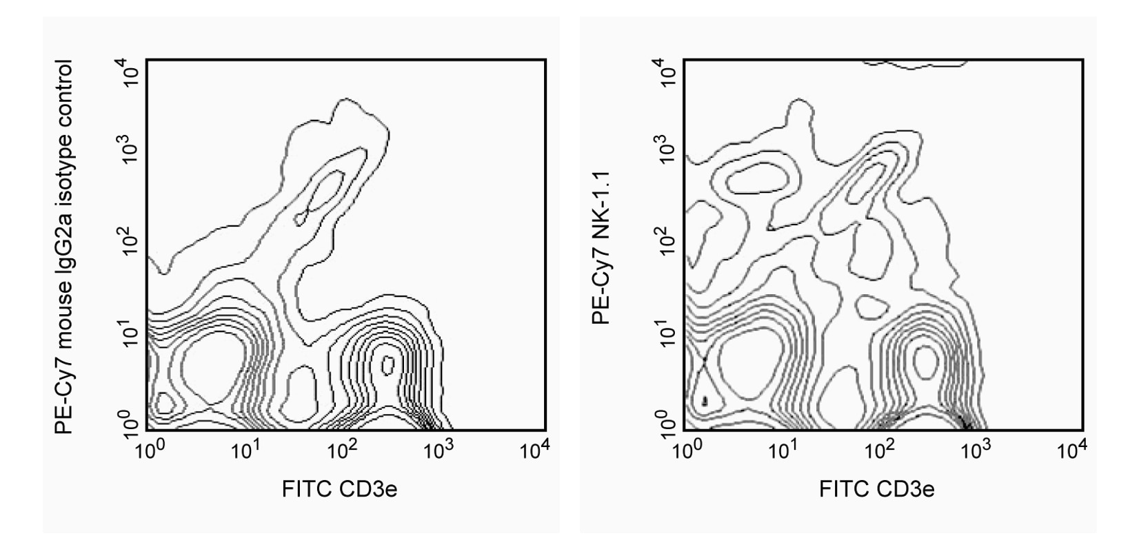
Two-color analysis of NK-1.1 expression on splenocytes. C57BL/6 splenocytes were stained with FITC-conjugated anti-mouse CD3e mAb 145-2C11 (Cat. No. 553061/553062) and either PE-Cy7-conjugated Mouse IgG2a κ isotype control mAb G155-178 (Cat. No. 552868, left panel) or PE-Cy7-conjugated mAb PK136 (right panel). NK-1.1+ CD3e- NK cells and NK-1.1[dim] CD3e+ NK-T cells are detected. Please note that the dead leukocytes were not excluded in this experiment, and the typical diagonal dead-cell population is partially obscured in the right panel. Flow cytometry was performed on a BD FACSCalibur™ Flow Cytometry System.
声明 :本官网所有报价均为常温或者蓝冰运输价格,如有产品需要干冰运输,需另外加收干冰运输费。