


 全部商品分类
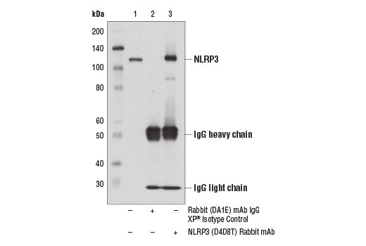


NLRP3 (D4D8T) Rabbit mAb


 下载产品说明书 下载COA 下载SDS
 用小程序,查商品更便捷


 收藏
收藏
 对比
对比 咨询
咨询
Monoclonal antibody is produced by immunizing animals with a synthetic peptide corresponding to residues surrounding Ala306 of mouse NLRP3 protein.
Product Usage Information
| Application | Dilution |
|---|---|
| Western Blotting | 1:1000 |
| Simple Western™ | 1:10 - 1:50 |
| Immunoprecipitation | 1:200 |
Specificity/Sensitivity
Species Reactivity:
Human, Mouse
Supplied in 10 mM sodium HEPES (pH 7.5), 150 mM NaCl, 100 µg/ml BSA, 50% glycerol and less than 0.02% sodium azide. Store at –20°C. Do not aliquot the antibody.
参考图片
Immunoprecipitation of NLRP3 from J774A.1 cell extracts using Rabbit (DA1E) mAb IgG XP® Isotype Control #3900 (lane 2) or NLRP3 (D4D8T) Rabbit mAb (lane 3). Lane 1 is 10% input. Western blot analysis was performed using NLRP3 (D4D8T) Rabbit mAb.
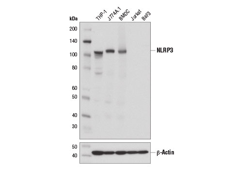
Western blot analysis of extracts from mouse bone marrow-derived dendritic cells (BMDC) and various cell lines using NLRP3 (D4D8T) Rabbit mAb (upper) and β-Actin (D6A8) Rabbit mAb #8457 (lower).
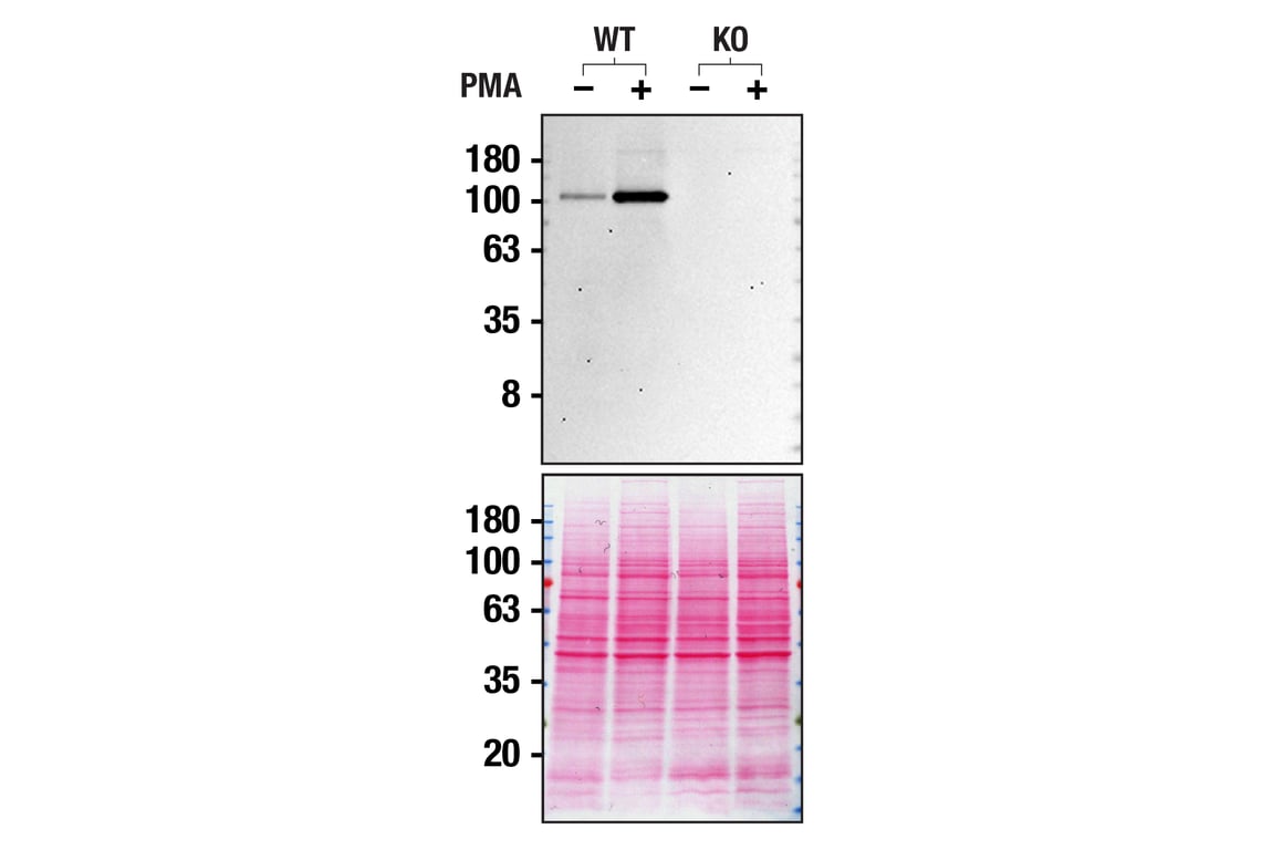
Western blot analysis of THP-1 extracts from WT or NLRP3 KO untreated (-) or treated with PMA (+) using NLRP3 (D4D8T) Rabbit mAb. Membranes stained with Ponceau S for total protein normalization (lower). These data were provided by YCharOS Inc., an open science company with the mission of characterizing commercially available antibodies, as a companion to validation data generated by CST scientists.
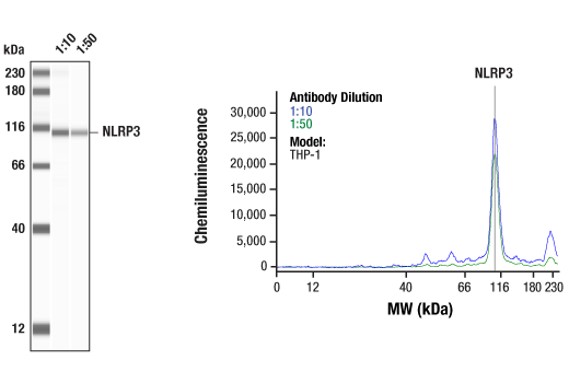
Simple Western™ analysis of lysates (0.1 mg/mL) from THP-1 cells using NLRP3 (D4D8T) Rabbit mAb #15101. The virtual lane view (left) shows the target band (as indicated) at 1:10 and 1:50 dilutions of primary antibody. The corresponding electropherogram view (right) plots chemiluminescence by molecular weight along the capillary at 1:10 (blue line) and 1:50 (green line) dilutions of primary antibody. This experiment was performed under reducing conditions on the Jess™ Simple Western instrument from ProteinSimple, a BioTechne brand, using the 12-230 kDa separation module.