


 全部商品分类


Non-Canonical BAF Complex Antibody Sampler Kit

 下载产品说明书 下载SDS
 用小程序,查商品更便捷


 收藏
收藏
 对比
对比 咨询
咨询The Non-Canonical BAF Complex Antibody Sampler Kit provides an economical means of detecting non-canonical BAF complex members using antibodies. The kit includes enough antibodies to perform two western blot experiments with each primary antibody.

参考图片
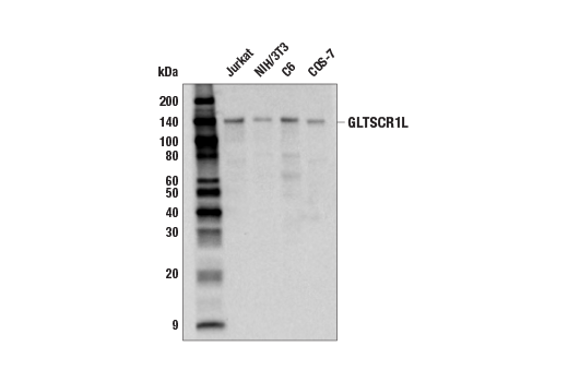
Western blot analysis of extracts from various cell lines using GLTSCR1L (E9K6F) Rabbit mAb.
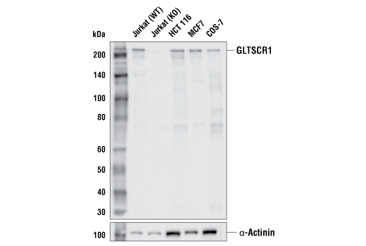
Western blot analysis of extracts from various cell lines using GLTSCR1 (E6I3A) Rabbit mAb (upper) or α-Actinin (D6F6) XP® Rabbit mAb #6487 (lower). GLTSCR1/GLTSCR1L double knockout (KO) cell lysates were isolated and provided by the Kadoch laboratory.
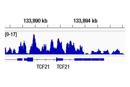
CUT&RUN was performed with NCCIT cells and GLTSCR1 (E6I3A) Rabbit mAb, using CUT&RUN Assay Kit #86652. DNA library was prepared using DNA Library Prep Kit for Illumina Systems (ChIP-seq, CUT&RUN) #56795. The figure shows binding across TCF21 gene.
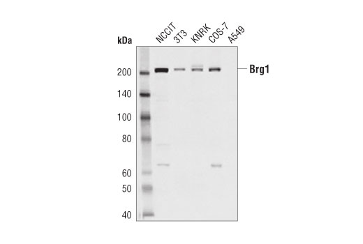
Western blot analysis of extracts from various cell lines using Brg1 (D1Q7F) Rabbit mAb.
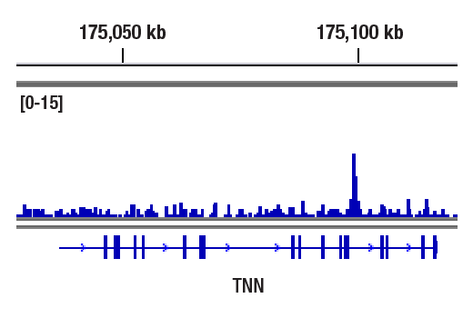
CUT&RUN was performed with NCCIT cells and Brg1 (D1Q7F) Rabbit mAb, using CUT&RUN Assay Kit #86652. DNA Library was prepared using DNA Library Prep Kit for Illumina® (ChIP-seq, CUT&RUN) #56795. The figure shows binding across TNN gene.
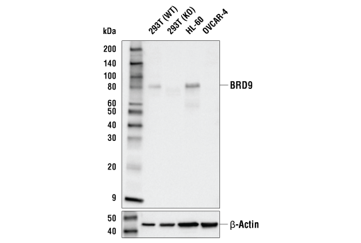
Western blot analysis of extracts from various cell lines using BRD9 (E9R2I) Rabbit mAb (upper) or β-Actin (D6A8) Rabbit mAb #8457 (lower). As expected, OVCAR-4 cells are low for BRD9 expression and no BRD9 is detected in the 293T knockout (KO) cells. BRD9 KO cell lysates were isolated and provided by the Kadoch laboratory.
After the primary antibody is bound to the target protein, a complex with HRP-linked secondary antibody is formed. The LumiGLO® is added and emits light during enzyme catalyzed decomposition.
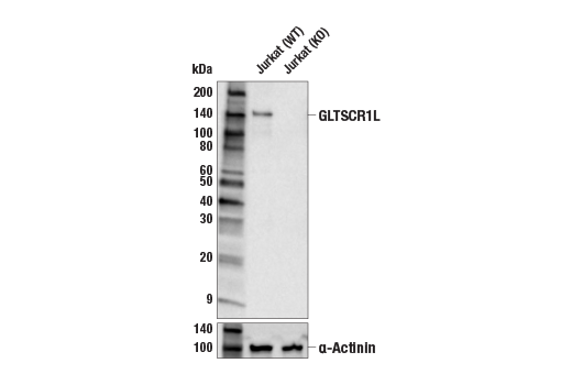
Western blot analysis of extracts from Jurkat cells, either wild-type (WT) or GLTSCR1/GLTSCR1L double knockout (KO), using GLTSCR1L (E9K6F) Rabbit mAb (upper) or α-Actinin (D6F6) XP® Rabbit mAb #6487 (lower). GLTSCR1/GLTSCR1L double KO cell lysates were isolated and provided by the Kadoch laboratory.
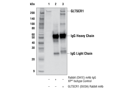
Immunoprecipitation of GLTSCR1 protein from Jurkat cell extracts. Lane 1 is 10% input, lane 2 is Rabbit (DA1E) mAb IgG XP® Isotype Control #3900, and lane 3 is GLTSCR1 (E6I3A) Rabbit mAb. Western blot analysis was performed using GLTSCR1 (E6I3A) Rabbit mAb. Anti-rabbit IgG, HRP-linked Antibody #7074 was used as a secondary antibody.
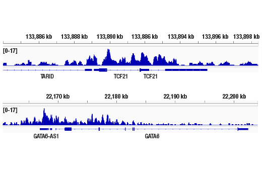
CUT&RUN was performed with NCCIT cells and GLTSCR1 (E6I3A) Rabbit mAb, using CUT&RUN Assay Kit #86652. DNA library was prepared using DNA Library Prep Kit for Illumina Systems (ChIP-seq, CUT&RUN) #56795. The figures show binding across TCF21 gene (upper) and GATA6 gene (lower).
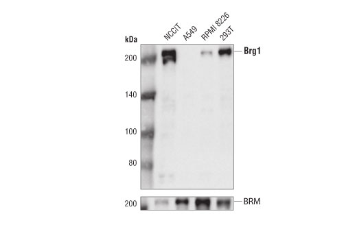
Western blot analysis of extracts from various cell lines using Brg1 (D1Q7F) Rabbit mAb (upper) or BRM (D9E8B) XP® Rabbit mAb (lower).
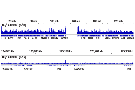
CUT&RUN was performed with NCCIT cells and Brg1 (D1Q7F) Rabbit mAb, using CUT&RUN Assay Kit #86652. DNA Library was prepared using DNA Library Prep Kit for Illumina® (ChIP-seq, CUT&RUN) #56795. The figures show binding across chromosome 1 (upper), including TNN gene (lower).
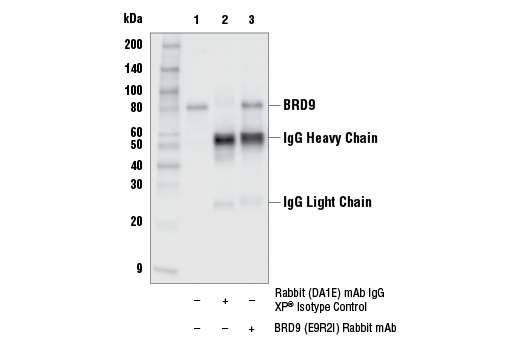
Immunoprecipitation of BRD9 protein from HDLM-2 cell extracts. Lane 1 is 10% input, lane 2 is Rabbit (DA1E) mAb IgG XP® Isotype Control #3900, and lane 3 is BRD9 (E9R2I) Rabbit mAb. Western blot analysis was performed using BRD9 (E9R2I) Rabbit mAb. Anti-rabbit IgG, HRP-linked Antibody #7074 was used as a secondary antibody.
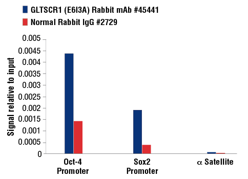
Chromatin immunoprecipitations were performed with cross-linked chromatin from untreated NCCIT cells and either GLTSCR1 (E6I3A) Rabbit mAb or Normal Rabbit IgG #2729 using SimpleChIP® Plus Enzymatic Chromatin IP Kit (Magnetic Beads) #9005. The enriched DNA was quantified by real-time PCR using SimpleChIP® Human Oct-4 Promoter Primers #4641, SimpleChIP® Human Sox2 Promoter Primers #4649, and SimpleChIP® Human α Satellite Repeat Primers #4486. The amount of immunoprecipitated DNA in each sample is represented as signal relative to the total amount of input chromatin, which is equivalent to one.
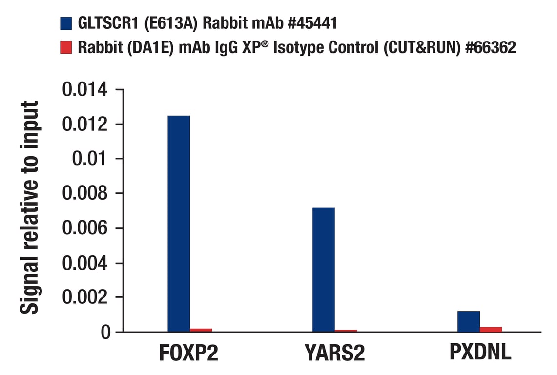
CUT&RUN was performed with NCCIT cells and either GLTSCR1 (E6I3A) Rabbit mAb or Rabbit (DA1E) mAb IgG XP® Isotype Control (CUT&RUN) #66362, using CUT&RUN Assay Kit #86652. The enriched DNA was quantified by real-time PCR using human FOXP2 promoter primers, human YARS2 promoter primers, and human PXDNL promoter primers. The amount of immunoprecipitated DNA in each sample is represented as signal relative to the total amount of input chromatin, which is equivalent to one.
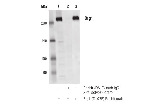
Immunoprecipitation of Brg1 from NCCIT cell extracts. Lane 1 is 10% input, lane 2 is Rabbit (DA1E) mAb IgG XP® Isotype Control #3900, and lane 3 is Brg1 (D1Q7F) Rabbit mAb. Western blot analysis was performed using Brg1 (D1Q7F) Rabbit mAb.
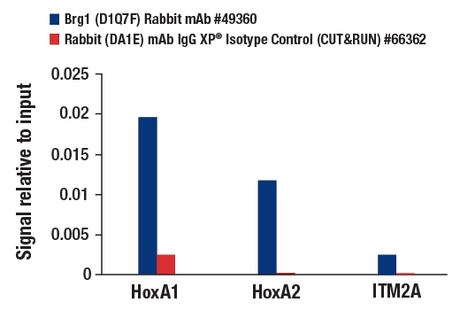
CUT&RUN was performed with NCCIT cells and either Brg1 (D1Q7F) Rabbit mAb or Rabbit (DA1E) mAb IgG XP® Isotype Control (CUT&RUN) #66362, using CUT&RUN Assay Kit #86652. The enriched DNA was quantified by real-time PCR using SimpleChIP® Human HoxA1 Intron 1 Primers #7707, SimpleChIP® Human HoxA2 Promoter Primers #5517, and human ITM2A upstream primers. The amount of immunoprecipitated DNA in each sample is represented as signal relative to the total amount of input chromatin, which is equivalent to one.
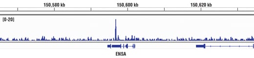
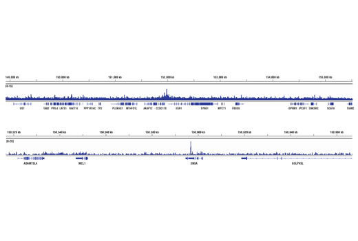
Chromatin immunoprecipitations were performed with cross-linked chromatin from MCF7 cells grown in phenol red free medium and 5% charcoal stripped FBS for 4 d followed by treatment with β-estradiol (10 nM, 45 min) and Brg1 (D1Q7F) XP® Rabbit mAb, using SimpleChIP® Enzymatic Chromatin IP Kit (Magnetic Beads) #9003. DNA Libraries were prepared using DNA Library Prep Kit for Illumina® (ChIP-seq, CUT&RUN) #56795. The figure shows binding across the ENSA gene.
Chromatin immunoprecipitations were performed with cross-linked chromatin from MCF7 cells grown in phenol red free medium and 5% charcoal stripped FBS for 4 d followed by treatment with β-estradiol (10 nM, 45 min) and Brg1 (D1Q7F) XP® Rabbit mAb, using SimpleChIP® Enzymatic Chromatin IP Kit (Magnetic Beads) #9003. DNA Libraries were prepared using DNA Library Prep Kit for Illumina® (ChIP-seq, CUT&RUN) #56795. The figure shows binding across ESR1 (upper), a known target gene of Brg1 (see additional figure containing ChIP-qPCR data), and ENSA gene (lower).
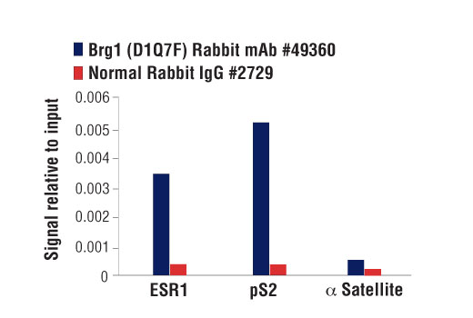
Chromatin immunoprecipitations were performed with cross-linked chromatin from MCF7 cells grown in phenol red free medium and 5% charcoal stripped FBS for 4 d followed by treatment with β-estradiol (10 nM, 45 min) and either Brg1 (D1Q7F) XP® Rabbit mAb or Normal Rabbit IgG #2729 using SimpleChIP® Enzymatic Chromatin IP Kit (Magnetic Beads) #9003. The enriched DNA was quantified by real-time PCR using SimpleChIP® Human ESR1 Promoter Primers #9673, SimpleChIP® Human pS2 Promoter Primers #9702, and SimpleChIP® Human α Satellite Repeat Primers #4486. The amount of immunoprecipitated DNA in each sample is represented as signal relative to the total amount of input chromatin, which is equivalent to one.
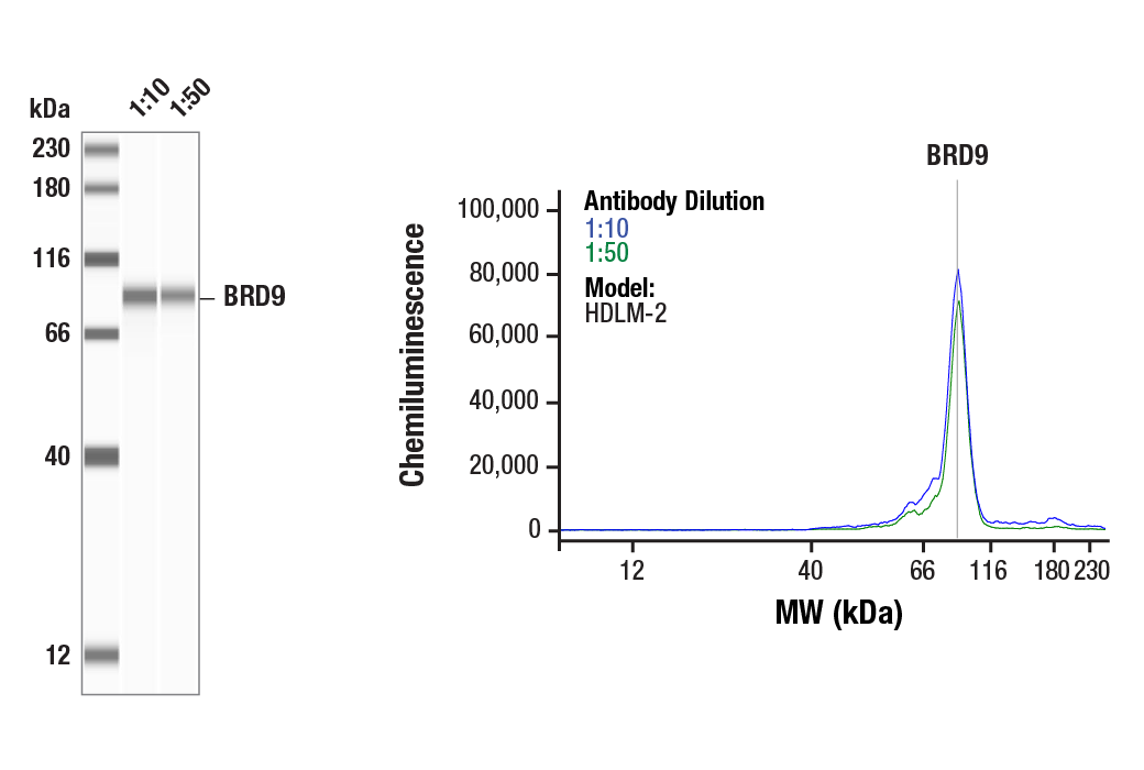
Simple Western™ analysis of lysates (1.0 mg/mL) from HDLM-2 cells using BRD9 (E9R2I) Rabbit mAb #58906. The virtual lane view (left) shows the target band (as indicated) at 1:10 and 1:50 dilutions of primary antibody. The corresponding electropherogram view (right) plots chemiluminescence by molecular weight along the capillary at 1:10 (blue line) and 1:50 (green line) dilutions of primary antibody. This experiment was performed under reducing conditions on the Jess™ Simple Western instrument from ProteinSimple, a BioTechne brand, using the 12-230 kDa separation module.
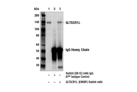
Immunoprecipitation of GLTSCR1L protein from Jurkat cell extracts. Lane 1 is 10% input, lane 2 is Rabbit (DA1E) mAb IgG XP® Isotype Control #3900, and lane 3 is GLTSCR1L (E9K6F) Rabbit mAb. Western blot analysis was performed using GLTSCR1L (E9K6F) Rabbit mAb. Anti-rabbit IgG, HRP-linked Antibody #7074 was used as a secondary antibody.