


 全部商品分类
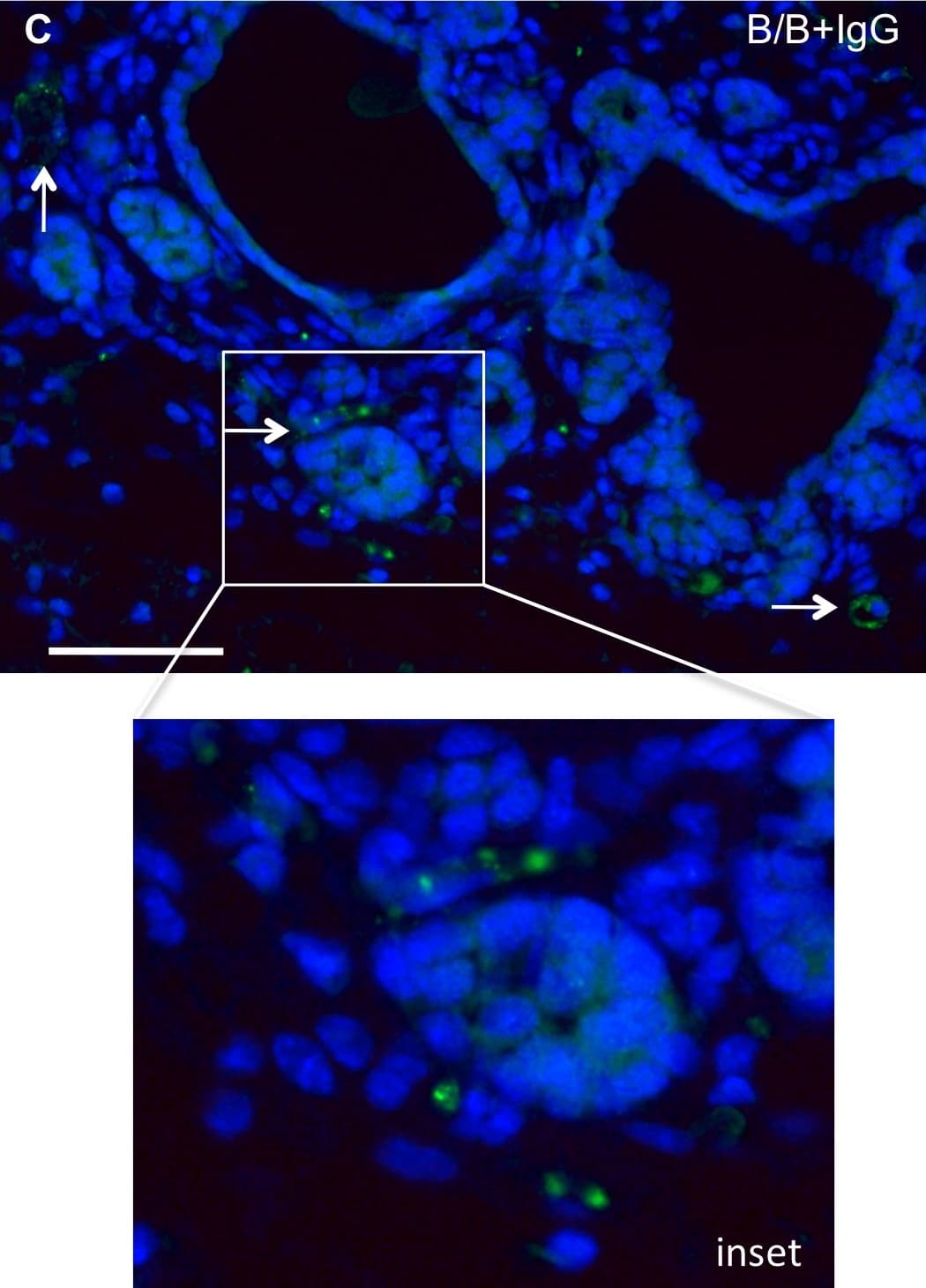


Normal Goat IgG Cont (1 mg)


 下载产品说明书 下载SDS
 用小程序,查商品更便捷


 收藏
收藏
 对比
对比 咨询
咨询
Scientific Data
 View Larger
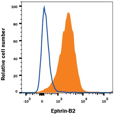
Detection of Normal Goat IgG Control by Flow Cytometry SH-SY5Y human neuroblastoma cell line was stained with Goat Anti-Human/Mouse/Rat Ephrin-B2 Antigen Affinity-purified Polyclonal Antibody (Catalog # AF496, filled histogram) or Normal Goat IgG Isotype Control Antibody (Catalog # AB-108-C, open histogram), followed by Phycoerythrin-conjugated Anti-Goat IgG Secondary Antibody (Catalog # F0107).
 View Larger
Detection of Mouse IgG by Immunocytochemistry/ Immunofluorescence Decreased macrophage recruitment correlates with decreased angiogenesis.A) Quantification of BrdU incorporation demonstrates that decreased macrophage infiltration does not significantly correlate with a change in epithelial cell proliferation (N.S. = not significant). B) Quantification of VWF staining reveals decreased blood vessels associated with epithelial structures in mammary glands from mice treated with B/B and anti-CX3CR1 blocking antibody compared with mice treated with B/B and IgG control antibody. *p<0.05. C, D) Representative images of VWF stained mammary glands from mice treated with B/B and either control IgG antibody (C) or anti-CX3CR1 (D). Green = VWF staining, blue = DAPI. Arrows indicate VWF-positive structures. Scale bars represent 50 µM. Results in each figure panel are representative of a minimum of three different mice for each treatment group and genotype. Image collected and cropped by CiteAb from the following open publication (https://dx.plos.org/10.1371/journal.pone.0045877), licensed under a CC-BY license. Not internally tested by R&D Systems.
 View Larger
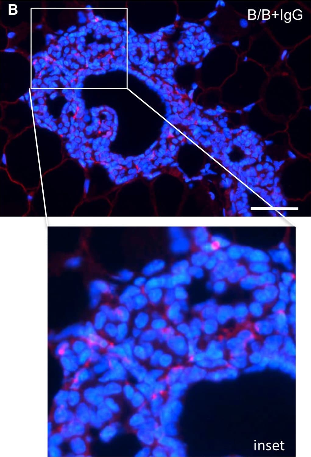
Detection of Mouse IgG by Immunocytochemistry/ Immunofluorescence iFGFR1 activation in vivo promotes recruitment of CX3CR1-positive macrophages.MMTV-iFGFR1 transgenic mice were treated with B/B in order to analyze the population of macrophages that are recruited to the mammary epithelium during early stages of iFGFR1-induced mammary tumorigenesis. A) MMTV-iFGFR1 mice treated with B/B demonstrated an increase in macrophage recruitment after 10 days as indicated by an increased in the number of F4/80 positive cells. MMTV-iFGFR1 mice treated with anti-CX3CR1 in conjunction with B/B demonstrated a reduction in macrophage recruitment at 10 days indicating that iFGFR1 activation is responsible for recruiting a subset of macrophages that express CX3CR1. ***p<0.0001. Error bars represent SEM. B) Representative image of macrophages associated with budding epithelial structures in mammary glands from mice treated with control IgG antibody. C) Representative image of macrophages associated with budding epithelial structures in mammary glands from mice treated with anti-CX3CR1. Red = F4/80 staining, blue = DAPI. Scale bars represent 50 µM. Results in each figure panel are representative of a minimum of three different mice for each treatment group and genotype. Image collected and cropped by CiteAb from the following open publication (https://dx.plos.org/10.1371/journal.pone.0045877), licensed under a CC-BY license. Not internally tested by R&D Systems.
 View Larger
Detection of Normal Goat IgG Control by Immunocytochemistry/ Immunofluorescence iFGFR1 activation in vivo promotes recruitment of CX3CR1-positive macrophages.MMTV-iFGFR1 transgenic mice were treated with B/B in order to analyze the population of macrophages that are recruited to the mammary epithelium during early stages of iFGFR1-induced mammary tumorigenesis. A) MMTV-iFGFR1 mice treated with B/B demonstrated an increase in macrophage recruitment after 10 days as indicated by an increased in the number of F4/80 positive cells. MMTV-iFGFR1 mice treated with anti-CX3CR1 in conjunction with B/B demonstrated a reduction in macrophage recruitment at 10 days indicating that iFGFR1 activation is responsible for recruiting a subset of macrophages that express CX3CR1. ***p<0.0001. Error bars represent SEM. B) Representative image of macrophages associated with budding epithelial structures in mammary glands from mice treated with control IgG antibody. C) Representative image of macrophages associated with budding epithelial structures in mammary glands from mice treated with anti-CX3CR1. Red = F4/80 staining, blue = DAPI. Scale bars represent 50 µM. Results in each figure panel are representative of a minimum of three different mice for each treatment group and genotype. Image collected and cropped by CiteAb from the following open publication (https://dx.plos.org/10.1371/journal.pone.0045877), licensed under a CC-BY license. Not internally tested by R&D Systems.
Background: IgG
R&D Systems offers a range of secondary antibodies and controls for flow cytometry, immunohistochemistry, and Western blotting. We provide species-specific secondary antibodies that are available with a variety of conjugated labels.
Our NorthernLights fluorescent secondary antibodies are bright and resistant to photobleaching. We are currently offering secondary antibodies recognizing mouse, rat, goat, sheep, and rabbit IgG as well as chicken IgY. These reagents are available with three distinct excitation and emission maxima, making them ideal for multi-color fluorescence microscopy.
Preparation and Storage
- 12 months from date of receipt, -20 to -70 °C as supplied.
- 1 month, 2 to 8 °C under sterile conditions after reconstitution.
- 6 months, -20 to -70 °C under sterile conditions after reconstitution.
参考图片
Detection of NormalGoat IgG Control by Flow Cytometry SH-SY5Y human neuroblastoma cell line was stained with Goat Anti-Human/Mouse/RatEphrin-B2 Antigen Affinity-purified Polyclonal Antibody (Catalog #AF496,filled histogram) or Normal Goat IgG Isotype Control Antibody(Catalog # AB-108-C, open histogram), followed by Phycoerythrin-conjugatedAnti-Goat IgG Secondary Antibody
(Catalog #F0107).