


 全部商品分类
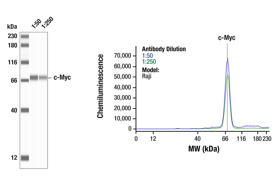


Notch Activated Targets Antibody Sampler Kit

 下载产品说明书 下载SDS
 用小程序,查商品更便捷


 收藏
收藏
 对比
对比 咨询
咨询
The Notch Activated Targets Antibody Sampler Kit provides an economical means of detecting target proteins of activated Notch. The kit contains enough primary antibody to perform four western blot experiments per primary antibody.
参考图片
Simple Western™ analysis of lysates (1 mg/mL) from Raji cells using c-Myc (D84C12) Rabbit mAb #5605. The virtual lane view (left) shows a single target band (as indicated) at 1:50 and 1:250 dilutions of primary antibody. The corresponding electropherogram view (right) plots chemiluminescence by molecular weight along the capillary at 1:50 (blue line) and 1:250 (green line) dilutions of primary antibody. This experiment was performed under reducing conditions on the Jess™ Simple Western instrument from ProteinSimple, a BioTechne brand, using the 12-230 kDa separation module.
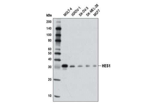
Western blot analysis of extracts from various cell lines using HES1 (D6P2U) Rabbit mAb.
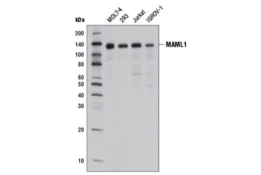
Western blot analysis of extracts from various cell lines using MAML1 (D3K7B) Rabbit mAb.
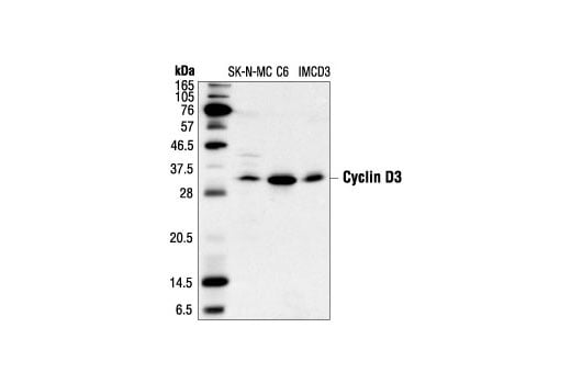
Western blot analysis of extracts from SK-N-MC, C6 and IMCD3 cells, using Cyclin D3 (DCS22) Mouse mAb.
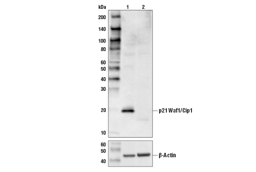
Western blot analysis from control HeLa cells (lane 1) or p21 Waf1/Cip1 knockout HeLa cells (lane 2) using p21 Waf1/Cip1 (12D1) Rabbit mAb (upper) or β-Actin (D6A8) Rabbit mAb #8457 (lower). The absence of signal in the p21 Waf1/Cip1 knockout HeLa cells confirms specificity of the antibody for p21 Waf1/Cip1.
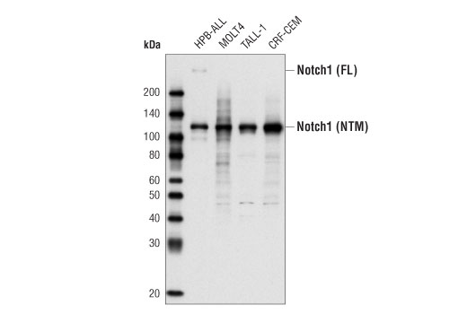
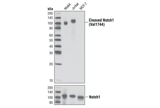
Western blot analysis of total cell extract from various cell types using Notch1 (D1E11) XP® Rabbit mAb. The full-length (FL) Notch protein and the cleaved transmembrane/intracellular region (NTM) are indicated.
After the primary antibody is bound to the target protein, a complex with HRP-linked secondary antibody is formed. The LumiGLO® is added and emits light during enzyme catalyzed decomposition.
After the primary antibody is bound to the target protein, a complex with HRP-linked secondary antibody is formed. The LumiGLO* is added and emits light during enzyme catalyzed decomposition.
Immunohistochemical analysis of paraffin-embedded human breast carcinoma, using Cyclin D3 (DCS22) Mouse mAb.
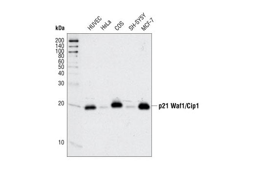
Western blot analysis of extracts from various cell types using p21 Waf1/Cip1 (12D1) Rabbit mAb.
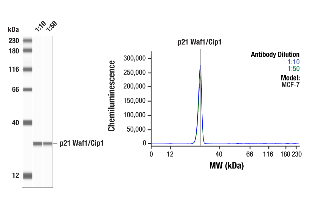
Simple Western™ analysis of lysates (1.0 mg/mL) from MCF-7 cells using p21 Waf1/Cip1 (12D1) Rabbit mAb #2947. The virtual lane view (left) shows a single target band (as indicated) at 1:10 and 1:50 dilutions of primary antibody. The corresponding electropherogram view (right) plots chemiluminescence by molecular weight along the capillary at 1:10 (blue line) and 1:50 (green line) dilutions of primary antibody. This experiment was performed under reducing conditions on the Jess™ Simple Western instrument from ProteinSimple, a BioTechne brand, using the 12-230 kDa separation module.
Western blot analysis of extracts from various cell lines using Cleaved Notch1 (Val1744) (D3B8) Rabbit mAb (upper) or Notch1 (D1E11) XP® Rabbit mAb #3608 (lower).
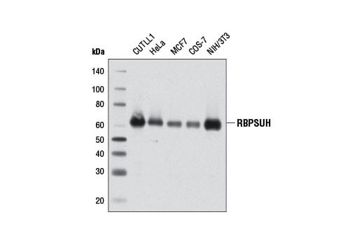
Western blot analysis of extracts from various cell lines using RBPSUH (D10A4) XP® Rabbit mAb.
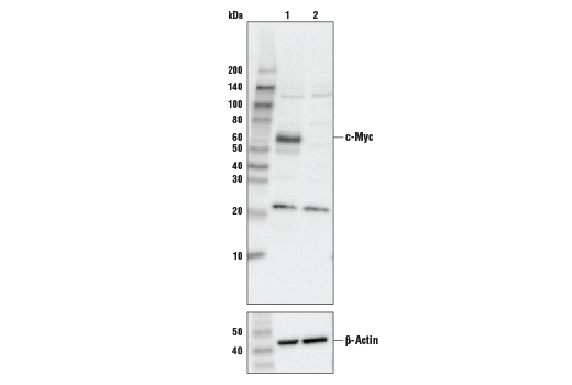
Western blot analysis of extracts from control HEK293 cells (lane 1) or c-Myc knockout HEK293 cells (lane 2) using c-Myc (D84C12) Rabbit mAb Antibody, #5605 (upper) or β-actin (13E5) Rabbit mAb, #4970 (lower). The absence of signal in the c-Myc knockout HEK293 cells confirms specificity of the antibody for c-Myc.
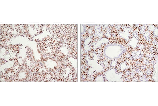
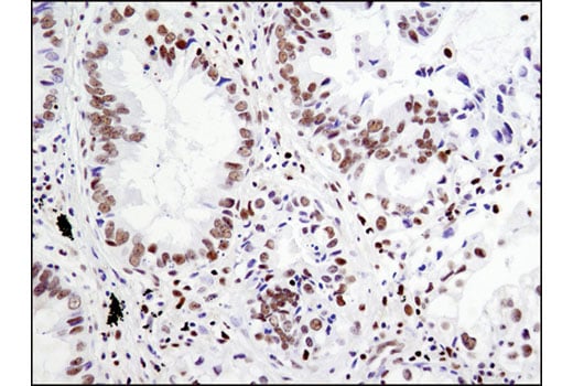
Immunohistochemical analysis of paraffin-embedded human lung carcinoma using HES1 (D6P2U) Rabbit mAb.
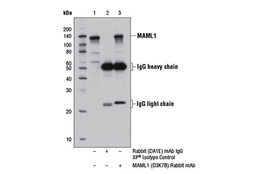
Immunoprecipitation of MAML1 protein from 293T cell extracts, using Rabbit (DA1E) mAb IgG XP® Isotype Control #3900 (lane 2) or MAML1 (D3K7B) Rabbit mAb (lane 3). Lane 1 is 10% input. Western blot analysis was performed using MAML1 Antibody #4608.
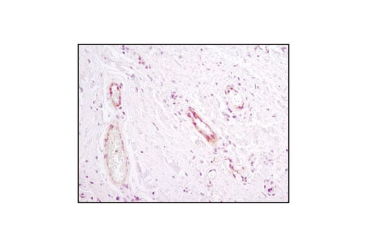
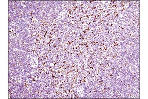
Immunohistochemical analysis of paraffin-embedded human inflammatory granulation tissue using Notch1 (D1E11) XP® Rabbit mAb.
Immunohistochemical analysis of paraffin-embedded human colon carcinoma using RBPSUH (D10A4) XP® Rabbit mAb.
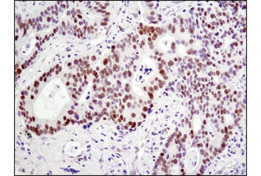
Immunohistochemical analysis of paraffin-embedded human breast carcinoma using HES1 (D6P2U) Rabbit mAb.
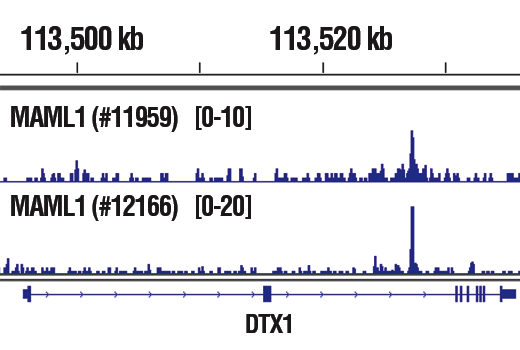
Chromatin immunoprecipitations were performed with cross-linked chromatin from CUTLL1 cells cultured in media with γ-secretase inhibitor (1 μM) for 3 days and then washed and cultured in fresh media for 3 hr and either MAML1 (D3E9) Rabbit mAb #11959 or MAML1 (D3K7B) Rabbit mAb, using SimpleChIP® Plus Enzymatic Chromatin IP Kit (Magnetic Beads) #9005. DNA Libraries were prepared using DNA Library Prep Kit for Illumina® (ChIP-seq, CUT&RUN) #56795. The figure shows binding across DTX1, a known target gene of MAML1 (see additional figure containing ChIP-qPCR data). For additional ChIP-seq tracks, please download the product datasheet.
Immunohistochemical analysis of paraffin-embedded human lung carcinoma, using Cyclin D3 (DCS22) Mouse mAb.
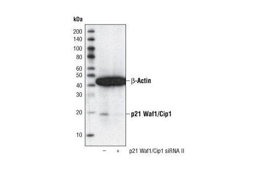
Western blot analysis of extracts from HeLa cells, transfected with 100 nM SignalSilence® Control siRNA (Fluorescein Conjugate) #6201 (-) or SignalSilence® p21 Waf1/Cip1 siRNA II (+), using p21 Waf1/Cip1 (12D1) Rabbit mAb #2947 and α-Tubulin (11H10) Rabbit mAb #2125. The p21 Waf1/Cip1 (12D1) Rabbit mAb confirms silencing of p21 Waf1/Cip1 expression and α-Tubulin (11H10) Rabbit mAb is used to control for loading and specificity of p21 Waf1/Cip1 siRNA.
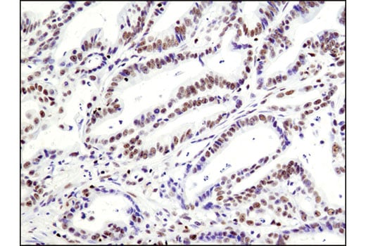
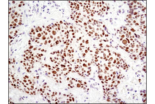
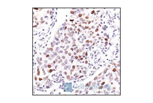
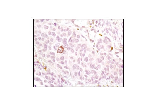
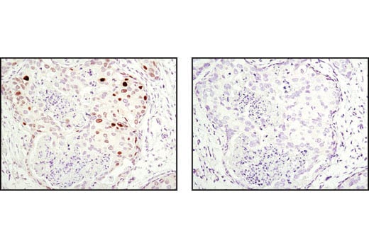
Immunohistochemical analysis of paraffin-embedded human breast carcinoma using Notch1 (D1E11) XP® Rabbit mAb.
Immunohistochemical analysis of paraffin-embedded human lung carcinoma using RBPSUH (D10A4) XP® Rabbit mAb in the presence of control peptide (left) or antigen-specific peptide (right).
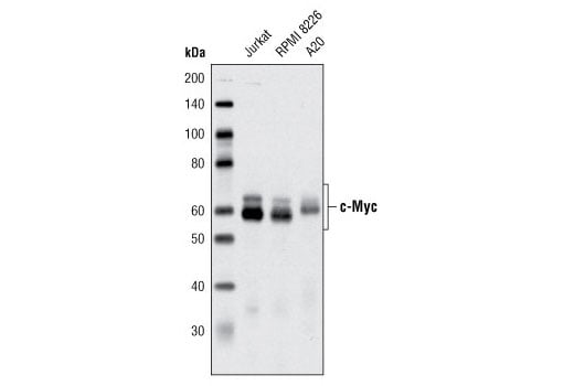
Western blot analysis of extracts from various cell lines using c-Myc (D84C12) Rabbit mAb.
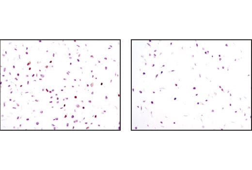
Immunohistochemical analysis of paraffin-embedded CUTLL1 cell pellets, control (left) or Compound E-treated (right), using HES1 (D6P2U) Rabbit mAb.
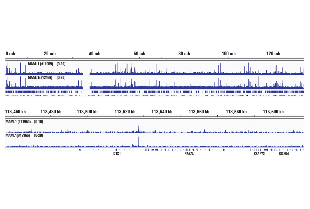
Chromatin immunoprecipitations were performed with cross-linked chromatin from CUTLL1 cells cultured in media with γ-secretase inhibitor (1 μM) for 3 days and then washed and cultured in fresh media for 3 hr and either MAML1 (D3E9) Rabbit mAb #11959 or MAML1 (D3K7B) Rabbit mAb, using SimpleChIP® Plus Enzymatic Chromatin IP Kit (Magnetic Beads) #9005. DNA Libraries were prepared using DNA Library Prep Kit for Illumina® (ChIP-seq, CUT&RUN) #56795. The figure shows binding across chromosome 12 (upper), including DTX1 (lower), a known target gene of MAML1 (see additional figure containing ChIP-qPCR data).
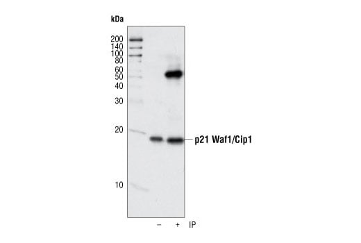
Immunoprecipitation of p21 from human umbillical vein endothelial cells (HUVECs) using p21 Waf1/Cip1 (12D1) Rabbit mAb. Western blot detection was performed using the same antibody.
Immunohistochemical analysis of paraffin-embedded A2780 (left), Jurkat (center) and RL (right) cell pellets using Notch1 (D1E11) XP® Rabbit mAb. Both A2780 and Jurkat express Notch1, but only Jurkat cells have cleaved Notch1, while RL cells express very low or no Notch1.
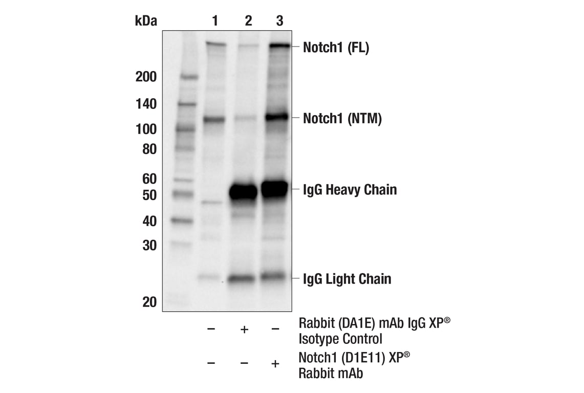
Immunoprecipitation of Notch1 protein from Molt-4 cell extracts. Lane 1 is 10% input, lane 2 is Rabbit (DA1E) mAb IgG XP® Isotype Control #3900, and lane 3 is Notch1 (D1E11) XP® Rabbit mAb. Western blot analysis was performed using Notch1 (D1E11) XP® Rabbit mAb.
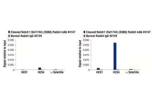
CUTLL1 cells were cultured in media with γ-secretase inhibitor (1μM) for 3 days and then either harvested immediately (left panel) or washed and cultured in fresh media for 3h (right panel). Chromatin immunoprecipitations were performed with cross-linked chromatin from cells and Cleaved Notch1 (Val1744) (D3B8) Rabbit mAb or Normal Rabbit IgG #2729 using SimpleChIP® Enzymatic Chromatin IP Kit (Magnetic Beads) #9003. The enriched DNA was quantified by real-time PCR using human HES1 promoter primers, SimpleChIP® Human HES4 Promoter Primers #7273, and SimpleChIP® Human α Satellite Repeat Primers #4486. The amount of immunoprecipitated DNA in each sample is represented as signal relative to the total amount of input chromatin, which is equivalent to one.
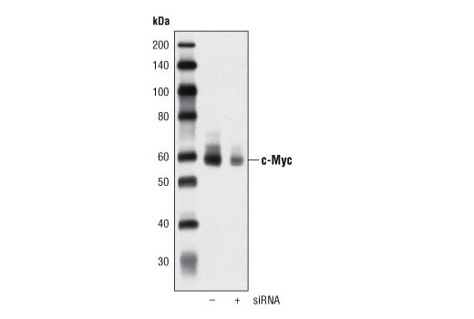
Western blot analysis of extracts from HeLa cells, mock transfected or transfected with SignalSilence® c-Myc siRNA I #6341, using c-Myc (D84C12) Rabbit mAb.
Immunohistochemical analysis of paraffin-embedded E18.5 mouse lung, Rbpjk F/+ Shh+/+ (wild type, left) or Rbpjk F/- Shhcre/+ (Rbpjk conditional knock out, right), using RBPSUH (D10A4) XP® Rabbit mAb. Note lack of staining in the bronchial epithelial cells in the conditional knock out tissue (right). Tissue courtesy of Dr. Wellington Cardosa, Boston University School of Medicine.
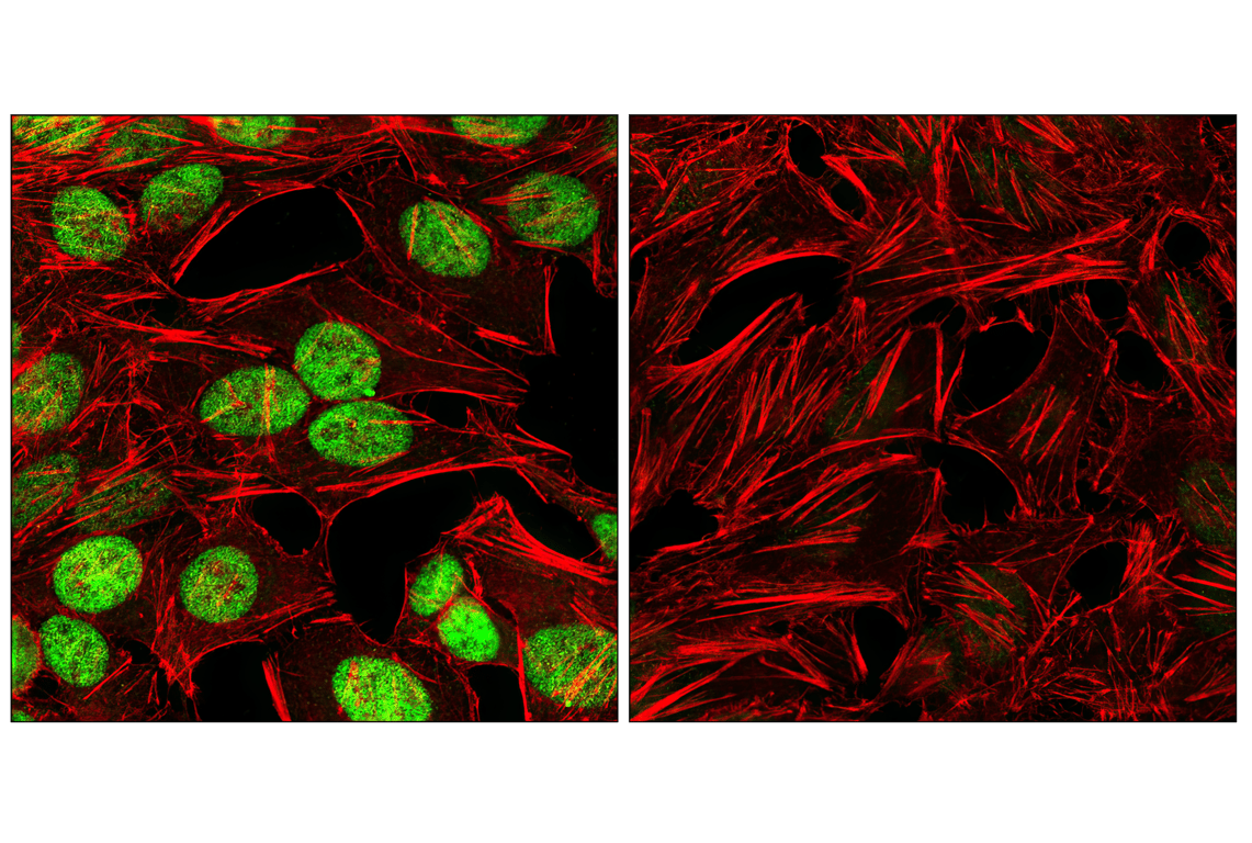
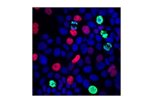
Confocal immunofluorescent analysis of HeLa cells, mock-transfected (left) or transfected with SignalSilence® c-Myc siRNA I #6341 (right), using c-Myc (D84C12) Rabbit mAb (green) and DyLight 554 Phalloidin #13054 (red).
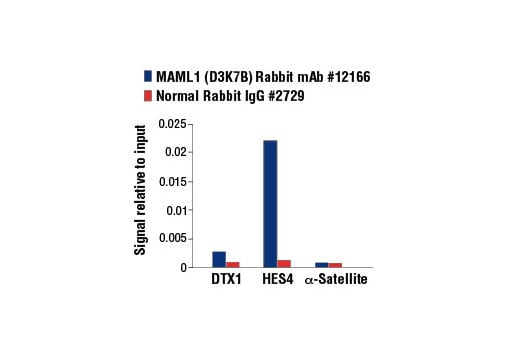
Chromatin immunoprecipitations were performed with cross-linked chromatin from CUTLL1 cells cultured in media with γ-secretase inhibitor (1 μM) for 3 days and then washed and cultured in fresh media for 3 hr and either MAML1 (D3K7B) Rabbit mAb or Normal Rabbit IgG #2729 using SimpleChIP® Enzymatic Chromatin IP Kit (Magnetic Beads) #9003. The enriched DNA was quantified by real-time PCR using human DTX1 intron 3 primers, SimpleChIP® Human HES4 Promoter Primers #7273, and SimpleChIP® Human α Satellite Repeat Primers #4486. The amount of immunoprecipitated DNA in each sample is represented as signal relative to the total amount of input chromatin, which is equivalent to one.
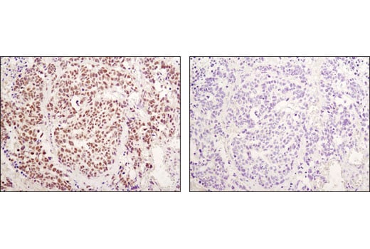
Immunohistochemical analysis of paraffin-embedded human breast carcinoma using p21 Waf1/Cip1 (12D1) Rabbit mAb in the presence of control peptide (left) or antigen-specific peptide (right).
Immunohistochemical analysis of paraffin-embedded human stomach adjacent to MALT (mucosa-associated lymphoid tissue) lymphoma using Notch1 (D1E11) XP® Rabbit mAb.
Immunohistochemical analysis of paraffin-embedded human lung carcinoma using RBPSUH (D10A4) XP® Rabbit mAb.
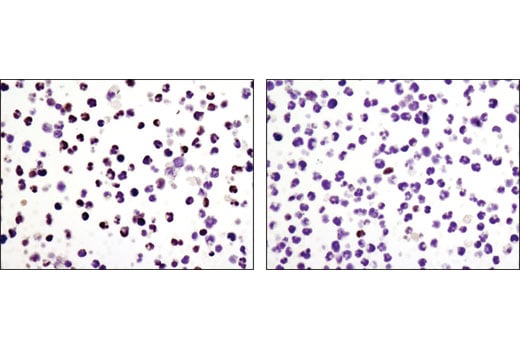
Immunohistochemical analysis of paraffin-embedded HeLa cells, transfected with SignalSilence® Control siRNA (Unconjugated) #6568 (left) or SignalSilence® p21 Waf1/Cip1 siRNA II #6558 (right), using p21 Waf1/Cip1 (12D1) Rabbit mAb.
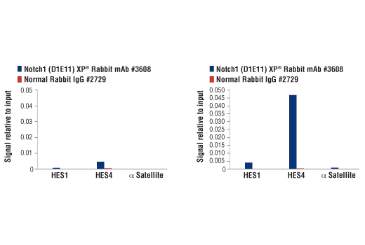
CUTLL1 cells were cultured in media with γ-secretase inhibitor (1 μM) for 3 days and then either harvested immediately (left panel) or washed and cultured in fresh media for 3 hr (right panel). Chromatin immunoprecipitations were performed with cross-linked chromatin from cells and Notch1 (D1E11) XP® Rabbit mAb or Normal Rabbit IgG #2729 using SimpleChIP® Enzymatic Chromatin IP Kit (Magnetic Beads) #9003. The enriched DNA was quantified by real-time PCR using human HES1 promoter primers, SimpleChIP® Human HES4 Promoter Primers #7273, and SimpleChIP® Human α Satellite Repeat Primers #4486. The amount of immunoprecipitated DNA in each sample is represented as signal relative to the total amount of input chromatin, which is equivalent to one.
Immunohistochemical analysis of paraffin-embedded mouse lymph node using RBPSUH (D10A4) XP® Rabbit mAb.
Confocal immunofluorescent analysis of MCF7 cells using p21 Waf1/Cip1 (12D1) Rabbit mAb (red) and Phospho-Histone H3 (Ser10) (6G3) Mouse mAb #9706 (green). Blue pseudocolor = DRAQ5® #4084 (fluorescent DNA dye).
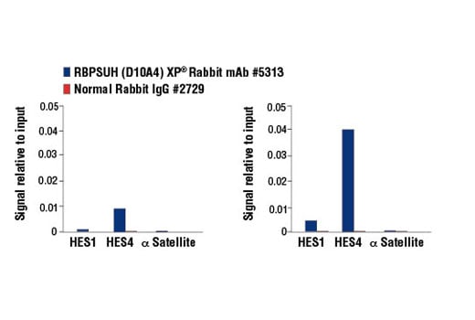
CUTLL1 cells were cultured in media with γ-secretase inhibitor (1 μM, 3 d) and then either harvested immediately (left panel) or washed and cultured in fresh media for 3 h (right panel). Chromatin immunoprecipitations were performed with cross-linked chromatin from cells and RBPSUH (D10A4) XP® Rabbit mAb or Normal Rabbit IgG #2729 using SimpleChIP® Enzymatic Chromatin IP Kit (Magnetic Beads) #9003. The enriched DNA was quantified by real-time PCR using human HES1 promoter primers, SimpleChIP® Human HES4 Promoter Primers #7273, and SimpleChIP® Human α Satellite Repeat Primers #4486. The amount of immunoprecipitated DNA in each sample is represented as signal relative to the total amount of input chromatin, which is equivalent to one.
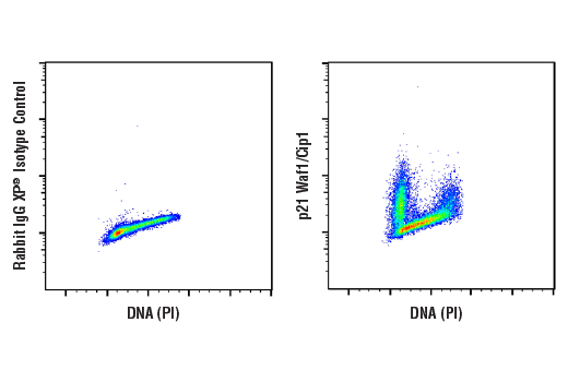
Flow cytometric analysis of Daudi cells using p21 Waf1/Cip1 (12D1) Rabbit mAb (right) and Propidium Iodide (PI)/RNase Staining Solution #4087, compared to concentration-matched Rabbit (DA1E) mAb IgG XP® Isotype Control #3900 (left). Anti-rabbit IgG (H+L), F(ab')2 Fragment (Alexa Fluor® 488 Conjugate) #4412 was used as a secondary antibody.