


 全部商品分类
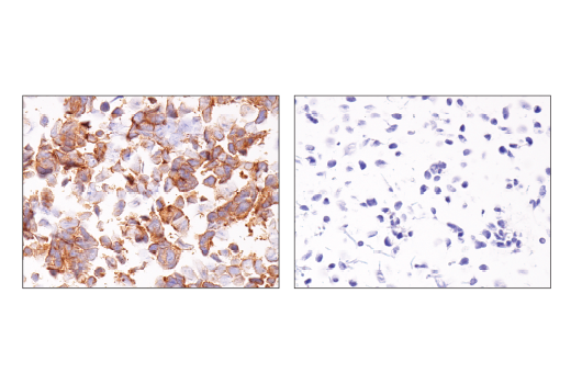


Notch Isoform Antibody Sampler Kit

 下载产品说明书 下载SDS
 用小程序,查商品更便捷


 收藏
收藏
 对比
对比 咨询
咨询
The Notch Isoform Antibody Sampler Kit provides an economical means to investigate Notch Signaling. The kit contains primary and secondary antibodies to perform two western mini-blots with each antibody.
参考图片
Immunohistochemical analysis of paraffin-embedded 293T cell pellet (left, positive) or IMR-32 cell pellet (right, negative) using Notch2 (D76A6) XP® Rabbit mAb.
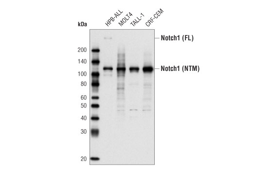
Western blot analysis of total cell extract from various cell types using Notch1 (D1E11) XP® Rabbit mAb. The full-length (FL) Notch protein and the cleaved transmembrane/intracellular region (NTM) are indicated.
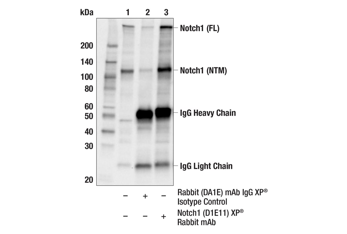
Immunoprecipitation of Notch1 protein from Molt-4 cell extracts. Lane 1 is 10% input, lane 2 is Rabbit (DA1E) mAb IgG XP® Isotype Control #3900, and lane 3 is Notch1 (D1E11) XP® Rabbit mAb. Western blot analysis was performed using Notch1 (D1E11) XP® Rabbit mAb.
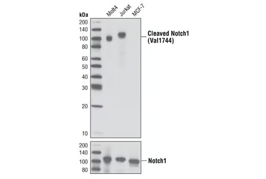
Western blot analysis of extracts from various cell lines using Cleaved Notch1 (Val1744) (D3B8) Rabbit mAb (upper) or Notch1 (D1E11) XP® Rabbit mAb #3608 (lower).
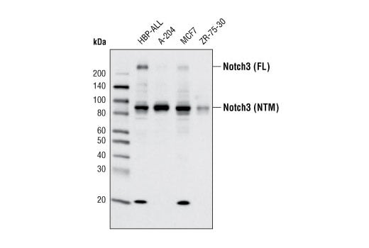
Western blot analysis of extracts from various cell lines using Notch3 (D11B8) Rabbit mAb.
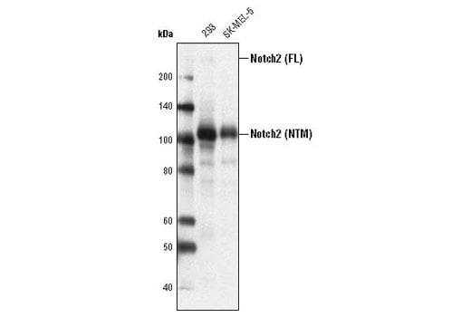
Western blot analysis of extracts from 293 and SK-MEL-5 cells using Notch2 (D76A6) XP® Rabbit mAb. The full-length (FL) Notch2 protein and the transmembrane/intracellular region (NTM) are indicated.
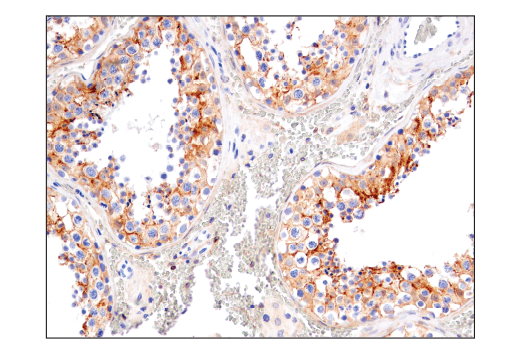
Immunohistochemical analysis of paraffin-embedded human serous papillary carcinoma of the ovary using Notch2 (D76A6) XP® Rabbit mAb performed on the Leica BOND RX.
After the primary antibody is bound to the target protein, a complex with HRP-linked secondary antibody is formed. The LumiGLO® is added and emits light during enzyme catalyzed decomposition.
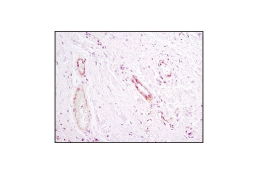
Immunohistochemical analysis of paraffin-embedded human inflammatory granulation tissue using Notch1 (D1E11) XP® Rabbit mAb.
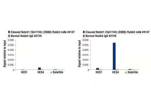
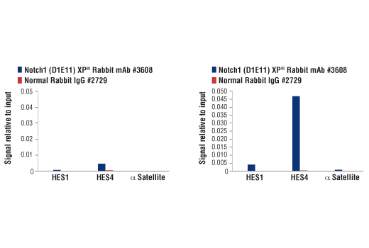
CUTLL1 cells were cultured in media with γ-secretase inhibitor (1μM) for 3 days and then either harvested immediately (left panel) or washed and cultured in fresh media for 3h (right panel). Chromatin immunoprecipitations were performed with cross-linked chromatin from cells and Cleaved Notch1 (Val1744) (D3B8) Rabbit mAb or Normal Rabbit IgG #2729 using SimpleChIP® Enzymatic Chromatin IP Kit (Magnetic Beads) #9003. The enriched DNA was quantified by real-time PCR using human HES1 promoter primers, SimpleChIP® Human HES4 Promoter Primers #7273, and SimpleChIP® Human α Satellite Repeat Primers #4486. The amount of immunoprecipitated DNA in each sample is represented as signal relative to the total amount of input chromatin, which is equivalent to one.
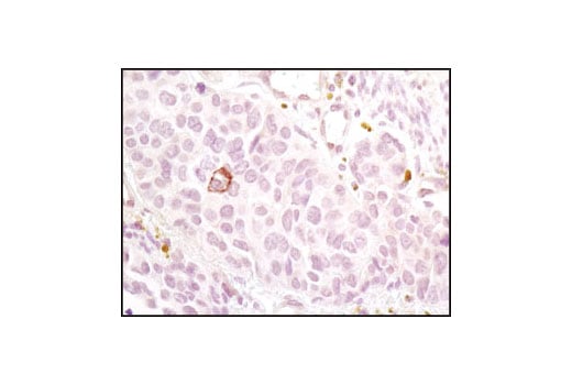
Immunohistochemical analysis of paraffin-embedded human papillary thyroid carcinoma using Notch2 (D76A6) XP® Rabbit mAb performed on the Leica BOND RX.
Immunohistochemical analysis of paraffin-embedded human breast carcinoma using Notch1 (D1E11) XP® Rabbit mAb.
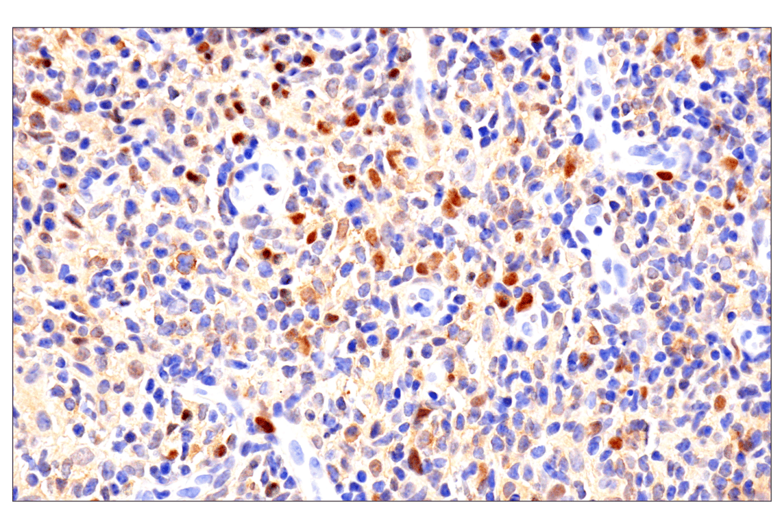
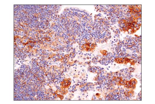
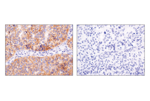
Immunohistochemical analysis of paraffin-embedded human non-Hodgkin lymphoma using Notch2 (D76A6) XP® Rabbit mAb performed on the Leica BOND RX.
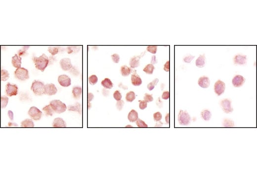
Immunohistochemical analysis of paraffin-embedded A2780 (left), Jurkat (center) and RL (right) cell pellets using Notch1 (D1E11) XP® Rabbit mAb. Both A2780 and Jurkat express Notch1, but only Jurkat cells have cleaved Notch1, while RL cells express very low or no Notch1.
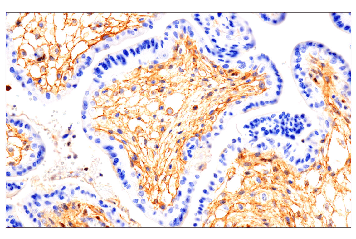
Immunohistochemical analysis of paraffin-embedded normal human placenta using Notch2 (D76A6) XP® Rabbit mAb performed on the Leica BOND RX.
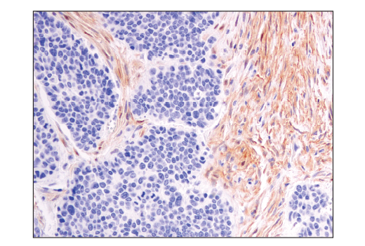
Immunohistochemical analysis of paraffin-embedded human stomach adjacent to MALT (mucosa-associated lymphoid tissue) lymphoma using Notch1 (D1E11) XP® Rabbit mAb.
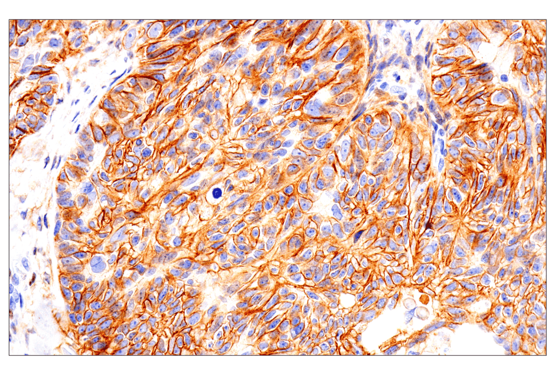
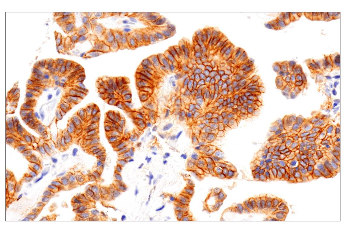
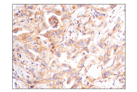
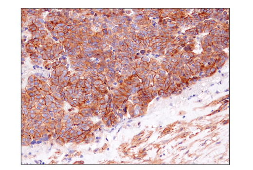
Immunohistochemical analysis of paraffin-embedded human ductal breast carcinoma using Notch2 (D76A6) XP® Rabbit mAb.
CUTLL1 cells were cultured in media with γ-secretase inhibitor (1 μM) for 3 days and then either harvested immediately (left panel) or washed and cultured in fresh media for 3 hr (right panel). Chromatin immunoprecipitations were performed with cross-linked chromatin from cells and Notch1 (D1E11) XP® Rabbit mAb or Normal Rabbit IgG #2729 using SimpleChIP® Enzymatic Chromatin IP Kit (Magnetic Beads) #9003. The enriched DNA was quantified by real-time PCR using human HES1 promoter primers, SimpleChIP® Human HES4 Promoter Primers #7273, and SimpleChIP® Human α Satellite Repeat Primers #4486. The amount of immunoprecipitated DNA in each sample is represented as signal relative to the total amount of input chromatin, which is equivalent to one.
Immunohistochemical analysis of paraffin-embedded human colon carcinoma using Notch2 (D76A6) XP® Rabbit mAb.
Immunohistochemical analysis of paraffin-embedded human B-cell non-Hodgkin's lymphoma using Notch2 (D76A6) XP® Rabbit mAb.
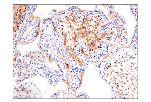
Immunohistochemical analysis of paraffin-embedded human ovarian serous carcinoma using Notch2 (D76A6) XP® Rabbit mAb.
Immunohistochemical analysis of paraffin-embedded human placenta using Notch2 (D76A6) XP® Rabbit mAb.
Immunohistochemical analysis of paraffin-embedded human testis using Notch2 (D76A6) XP® Rabbit mAb.
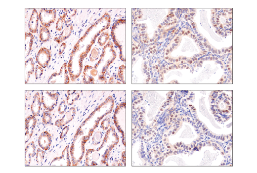
Immunohistochemical analysis of paraffin-embedded human prostate adenocarcinoma (left) or endometrioid adenocarcinoma (right) using Notch2 (D76A6) XP® Rabbit mAb (top) or Notch2 Rabbit mAb (bottom). These two antibodies detect independent, unique epitopes on human Notch2. Both epitopes are contained within the intracellular domain and the nuclear staining represents active Notch2. The similar staining patterns obtained with both antibodies help to confirm the specificity of the staining.
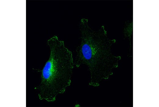
Confocal immunofluorescent analysis of SNB19 cells using Notch2 (D76A6) XP® Rabbit mAb (green). Blue pseudocolor = DRAQ5® #4084 (fluorescent DNA dye).
Immunohistochemical analysis of paraffin-embedded human serous papillary carcinoma of the ovary using Notch2 (D76A6) XP® Rabbit mAb (left) compared to concentration-matched Rabbit (DA1E) mAb IgG XP® Isotype Control #3900 (right).
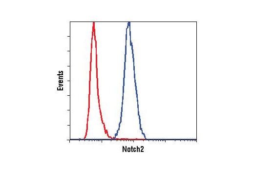
Flow cytometric analysis of fixed and permeabilized SW620 cells (red, low expression) and 293 cells (blue, positive) cells using Notch2 (D76A6) XP® Rabbit mAb. Anti-rabbit IgG (H+L), F(ab')2 Fragment (Alexa Fluor® 488 Conjugate) #4412 was used as a secondary antibody.