首页 抗体 一抗 一抗 NUT (C52B1) Rabbit mAb (Alexa Fluor ® 555 Conjugate)


1/1
产品介绍
产品信息
来源纯化
Monoclonal antibody is produced by immunizing animals with a recombinant protein corresponding to the human NUT protein.
简单描述
This Cell Signaling Technology antibody is conjugated to Alexa Fluor® 555 fluorescent dye and tested in-house for direct immunofluorescent analysis in rat tissue. This antibody is expected to exhibit the same species cross-reactivity as the unconjugated NUT (C52B1) Rabbit mAb #3625.
商品描述
Product Usage Information
| Application | Dilution |
|---|
| Immunofluorescence (Frozen) | 1:50 |
应用
目标/特异性
Specificity/Sensitivity
NUT (C52B1) Rabbit mAb (Alexa Fluor 555 Conjugate) detects endogenous levels of total NUT protein. The antibody also detects endogenous levels of the BRD4-NUT fusion protein found in NUT midline carcinoma (NMC).
Species Reactivity:
Human, Rat
背景
背景
Nuclear protein in testis (NUT) is normally confined to the germ cells of the testis and ovary (1,2). NUT midline carcinoma (NMC) is a recently recognized cancer that is defined by the presence of chromosomal rearrangements involving the NUT gene on chromosome 15q14 (3). In most cases the chromosomal translocation occurs between NUT and BRD4 on chromosome 19, resulting in the formation of a BRD4-NUT fusion protein. In the remaining tumors, variant NUT rearrangements are present involving BRD3, a very close homolog of BRD4. BRD4-NUT and BRD3-NUT encode fusion proteins that appear to contribute to carcinogenesis by blocking epithelial cell differentiation. NMCs, which are aggressive and highly lethal carcinomas, are morphologically indistinguishable from other poorly differentiated carcinomas. Given the limited expression of endogenous NUT protein, this antibody can be used to detect NUT fusion proteins in tissues by immunohistochemistry and immunofluorescence (2).
1.French, C.A. et al. (2003) Cancer Res 63, 304-7.
2.Haack, H. et al. (2009) Am J Surg Pathol 33, 984-91.
3.French, C.A. et al. (2008) Oncogene 27, 2237-42.

制备和贮存
保存方式
Supplied in PBS (pH 7.2), less than 0.1% sodium azide and 2 mg/ml BSA. Store at 4°C. Do not aliquot the antibody. Protect from light. Do not freeze.
参考图片
Confocal immunofluorescent analysis of rat testis (left, positive) or small intestine (right, negative) using NUT (C52B1) Rabbit mAb (Alexa Fluor® 555 Conjugate) (yellow). Actin filaments were labeled with DyLight™ 650 Phalloidin #12956 (red). Samples were mounted in ProLong® Gold Antifade Reagent with DAPI #8961 (blue).
当前规格1件起购
预计2-3周送达,快递: 免运费,若需干冰额外收费


 全部商品分类
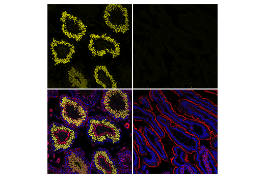

 下载产品说明书
 用小程序,查商品更便捷


 收藏
收藏
 对比
对比 咨询
咨询