

 全部商品分类
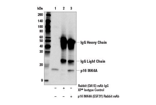


 用小程序,查商品更便捷


Monoclonal antibody is produced by immunizing animals with a synthetic peptide corresponding to residues surrounding Pro67 of mouse p16 INK4A protein.

Product Usage Information
| Application | Dilution |
|---|---|
| Western Blotting | 1:1000 |
| Immunoprecipitation | 1:100 |
Specificity/Sensitivity
Species Reactivity:
Mouse
Supplied in 10 mM sodium HEPES (pH 7.5), 150 mM NaCl, 100 µg/ml BSA, 50% glycerol and less than 0.02% sodium azide. Store at –20°C. Do not aliquot the antibody.
参考图片
Immunoprecipitation of p16 INK4A protein from mouse A20 cell extracts. Lane 1 is 10% input, lane 2 is Rabbit (DA1E) mAb IgG XP® Isotype Control #3900, and lane 3 is p16 INK4A (E5F3Y) Rabbit mAb. Western blot analysis was performed using p16 INK4A (E5F3Y) Rabbit mAb. Anti-rabbit IgG, HRP-linked Antibody #7074 was used as the secondary antibody.
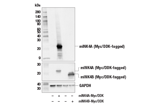
Western blot analysis of extracts from 293T cells, mock transfected (-) or transfected with constructs expressing full-length mouse p16 INK4A protein (mINK4A-Myc/DDK; +) or full-length mouse p15 INK4B protein (mINK4B-Myc/DDK; +), using p16 INK4A (E5F3Y) Rabbit mAb (upper), Myc-Tag (71D10) Rabbit mAb #2278 (middle), or GAPDH (D16H11) XP® Rabbit mAb #5174 (lower).
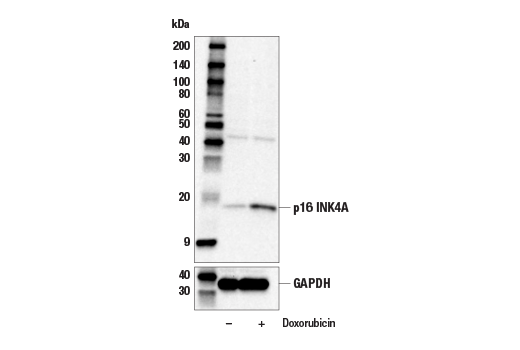
Western blot analysis of extracts from MEF cells, untreated (-) or treated with Doxorubicin #5927 (250 nM, 24 hours followed by 7 days without doxorubicin; +), using p16 INK4A (E5F3Y) Rabbit mAb (upper) or GAPDH (D16H11) XP® Rabbit mAb #5174 (lower). p16 INK4A protein expression is induced by doxorubicin as expected.
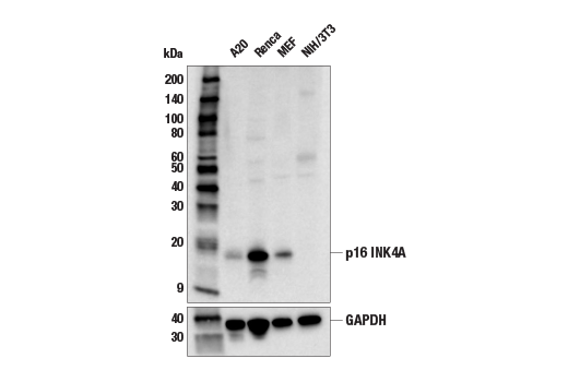
Western blot analysis of extracts from various mouse cell lines using p16 INK4A (E5F3Y) Rabbit mAb (upper) or GAPDH (D16H11) XP® Rabbit mAb #5174 (lower). Negative expression of p16 INK4A protein in NIH/3T3 cells is consistent with the predicted expression pattern.