


 全部商品分类


p21 Waf1/Cip1 (12D1) Rabbit mAb


 下载产品说明书 下载COA 下载SDS
 用小程序,查商品更便捷


 收藏
收藏
 对比
对比 咨询
咨询
Monoclonal antibody is produced by immunizing animals with a synthetic peptide corresponding to residues near the carboxy-terminus of human p21.
Product Usage Information
| Application | Dilution |
|---|---|
| Western Blotting | 1:1000 |
| Simple Western™ | 1:10 - 1:50 |
| Immunoprecipitation | 1:50 |
| Immunohistochemistry (Paraffin) | 1:50 |
| Immunofluorescence (Immunocytochemistry) | 1:400 - 1:800 |
| Flow Cytometry (Fixed/Permeabilized) | 1:100 - 1:400 |
Specificity/Sensitivity
Species Reactivity:
Human, Monkey
Supplied in 10 mM sodium HEPES (pH 7.5), 150 mM NaCl, 100 µg/ml BSA, 50% glycerol and less than 0.02% sodium azide. Store at –20°C. Do not aliquot the antibody.
For a carrier free (BSA and azide free) version of this product see product #19399.
参考图片
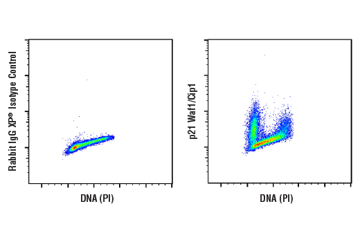
Flow cytometric analysis of Daudi cells using p21 Waf1/Cip1 (12D1) Rabbit mAb (right) and Propidium Iodide (PI)/RNase Staining Solution #4087, compared to concentration-matched Rabbit (DA1E) mAb IgG XP® Isotype Control #3900 (left). Anti-rabbit IgG (H+L), F(ab')2 Fragment (Alexa Fluor® 488 Conjugate) #4412 was used as a secondary antibody.
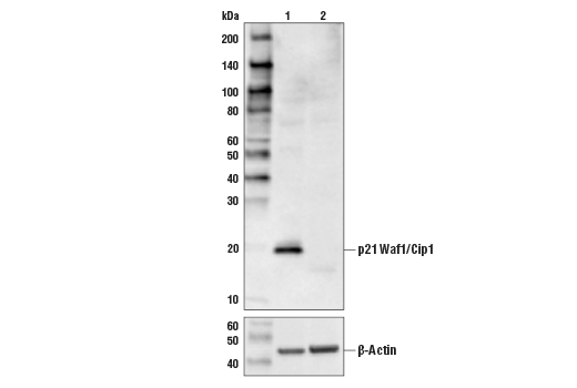
Western blot analysis from control HeLa cells (lane 1) or p21 Waf1/Cip1 knockout HeLa cells (lane 2) using p21 Waf1/Cip1 (12D1) Rabbit mAb (upper) or β-Actin (D6A8) Rabbit mAb #8457 (lower). The absence of signal in the p21 Waf1/Cip1 knockout HeLa cells confirms specificity of the antibody for p21 Waf1/Cip1.
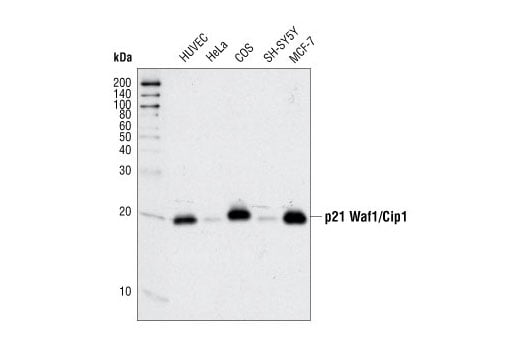
Western blot analysis of extracts from various cell types using p21 Waf1/Cip1 (12D1) Rabbit mAb.
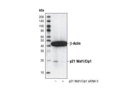
Western blot analysis of extracts from HeLa cells, transfected with 100 nM SignalSilence® Control siRNA (Fluorescein Conjugate) #6201 (-) or SignalSilence® p21 Waf1/Cip1 siRNA II (+), using p21 Waf1/Cip1 (12D1) Rabbit mAb #2947 and α-Tubulin (11H10) Rabbit mAb #2125. The p21 Waf1/Cip1 (12D1) Rabbit mAb confirms silencing of p21 Waf1/Cip1 expression and α-Tubulin (11H10) Rabbit mAb is used to control for loading and specificity of p21 Waf1/Cip1 siRNA.
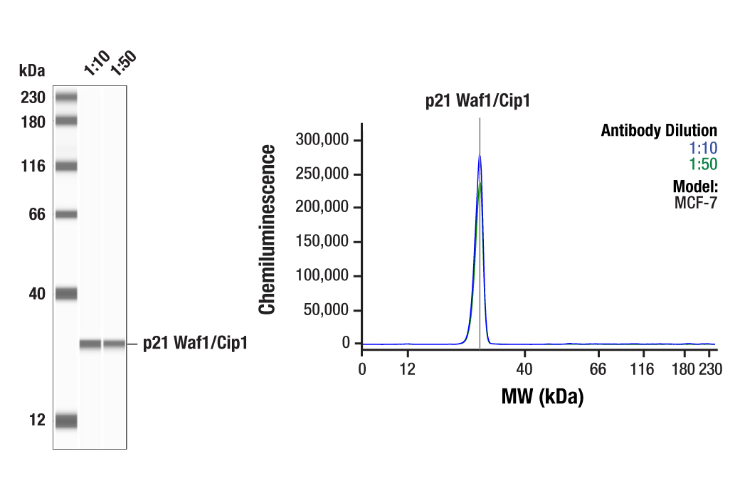
Simple Western™ analysis of lysates (1.0 mg/mL) from MCF-7 cells using p21 Waf1/Cip1 (12D1) Rabbit mAb #2947. The virtual lane view (left) shows a single target band (as indicated) at 1:10 and 1:50 dilutions of primary antibody. The corresponding electropherogram view (right) plots chemiluminescence by molecular weight along the capillary at 1:10 (blue line) and 1:50 (green line) dilutions of primary antibody. This experiment was performed under reducing conditions on the Jess™ Simple Western instrument from ProteinSimple, a BioTechne brand, using the 12-230 kDa separation module.
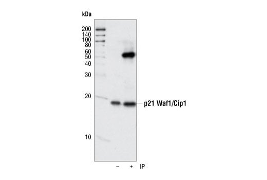
Immunoprecipitation of p21 from human umbillical vein endothelial cells (HUVECs) using p21 Waf1/Cip1 (12D1) Rabbit mAb. Western blot detection was performed using the same antibody.
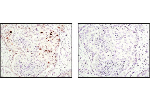
Immunohistochemical analysis of paraffin-embedded human breast carcinoma using p21 Waf1/Cip1 (12D1) Rabbit mAb in the presence of control peptide (left) or antigen-specific peptide (right).
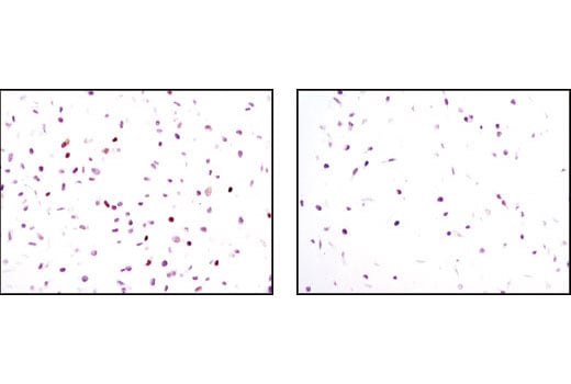
Immunohistochemical analysis of paraffin-embedded HeLa cells, transfected with SignalSilence® Control siRNA (Unconjugated) #6568 (left) or SignalSilence® p21 Waf1/Cip1 siRNA II #6558 (right), using p21 Waf1/Cip1 (12D1) Rabbit mAb.
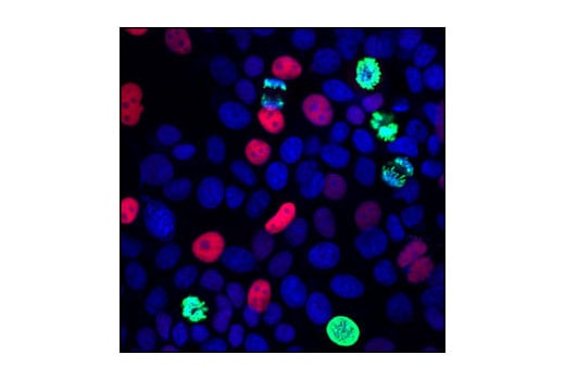
Confocal immunofluorescent analysis of MCF7 cells using p21 Waf1/Cip1 (12D1) Rabbit mAb (red) and Phospho-Histone H3 (Ser10) (6G3) Mouse mAb #9706 (green). Blue pseudocolor = DRAQ5® #4084 (fluorescent DNA dye).