


 下载产品说明书
 用小程序,查商品更便捷


 收藏
收藏
 对比
对比 咨询
咨询
Specificity/Sensitivity
参考图片
After the primary antibody is bound to the target protein, a complex with HRP-linked secondary antibody is formed. The LumiGLO* is added and emits light during enzyme catalyzed decomposition.
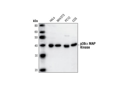
Western blot analysis of extracts from HeLa, NIH/3T3, PC12 and COS cells, using p38α MAPK Antibody.
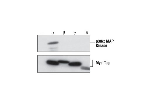
Western blot analysis of extracts from Xenopus oocytes overexpressing Myc-tagged p38 isoforms, using p38α MAPK Antibody (upper) and Myc-Tag Antibody (lower) to demonstrate the isoform specificity of p38α MAPK Antibody. (Provided by Dr. Eusebio Perdiguero and Dr. Angel Nebreda, European Molecular Biology Laboratory, Germany.)
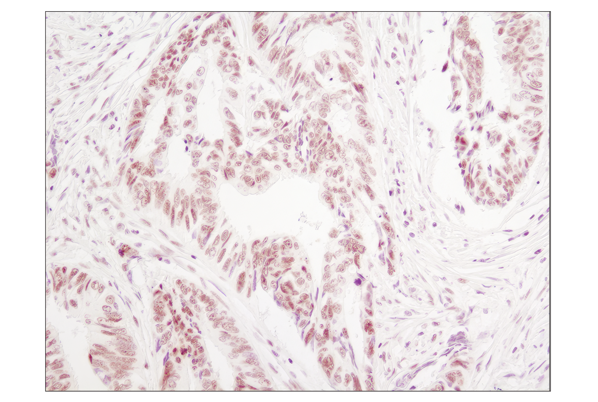
Immunohistochemical analysis of paraffin-embedded human breast carcinoma, showing cytoplasmic and nuclear localization, using p38α MAPK Antibody.
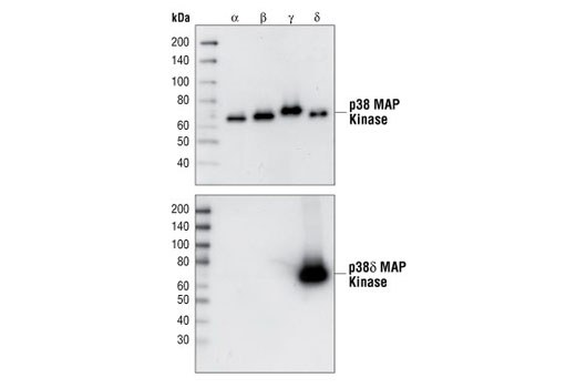
Western blot analysis of purified recombinant full-length p38 MAPK GST fusion protein, using p38 MAP kinase pan antibody (upper) or p38γ MAPK Antibody (lower).
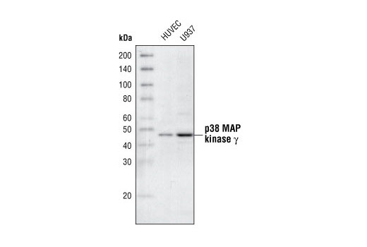
Western blot analysis of extracts from HUVEC and U937 cells, using p38γ MAPK Antibody.
Western blot analysis of extracts from 293, NBT-II and PC12 cells, using p38δ MAPK (10A8) Rabbit mAb.
Western blot analysis of purified recombinant full-length p38 MAPK GST fusion proteins, using p38 MAPK pan antibody (upper), or p38δ MAPK (10A8) Rabbit mAb (lower).
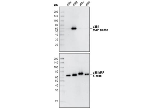
Western blot analysis of purified recombinant full-length p38 MAPK GST-fusion proteins, using p38β MAPK (C28C2) Rabbit mAb (upper), or a p38 MAPK pan antibody (lower).
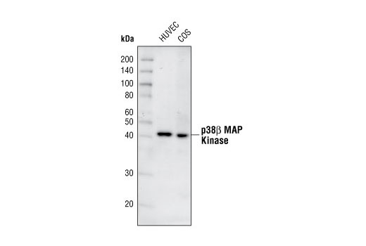
Westen blot analysis of extracts from HUVEC and COS cells using p38β MAPK (C28C2) Rabbit mAb.
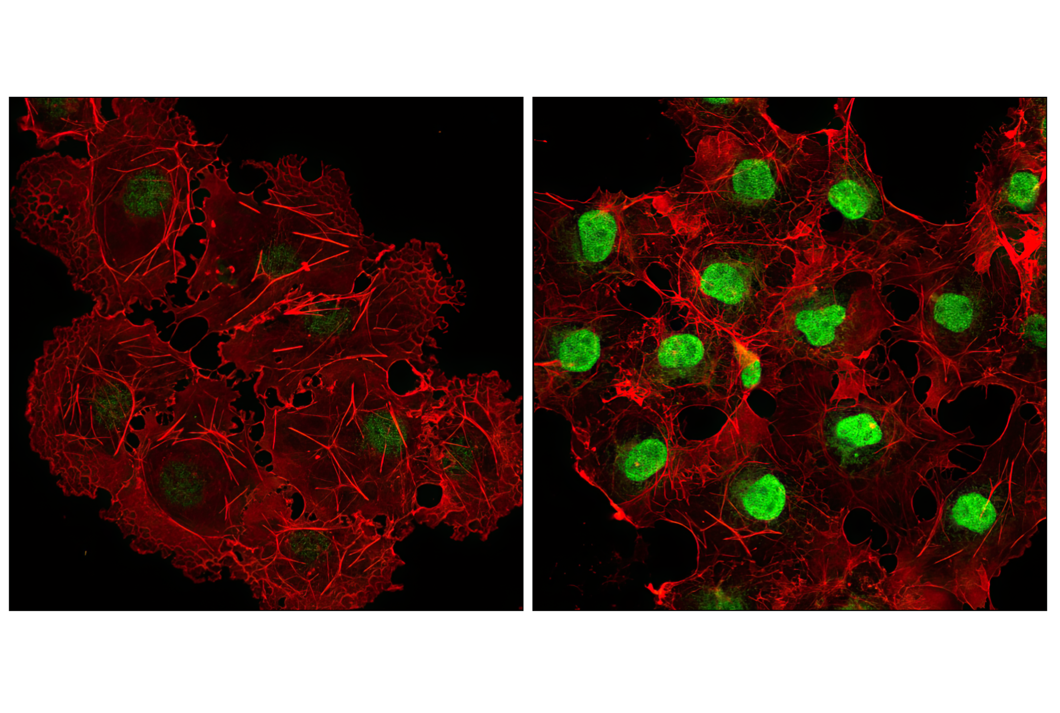
Confocal immunofluorescent analysis of COS cells, untreated (left) or anisomycin-treated (right) using Phospho-p38 MAPK (Thr180/Tyr182) (D3F9) XP® Rabbit mAb (green). Actin filaments have been labeled with DY-554 phalloidin (red).
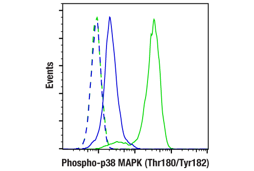
Flow cytometric analysis of Jurkat cells, untreated (blue) or anisomycin-treated (green), using Phospho-p38 MAPK (Thr180/Tyr182) (D3F9) XP® Rabbit mAb compared to a nonspecific negative control antibody (red).
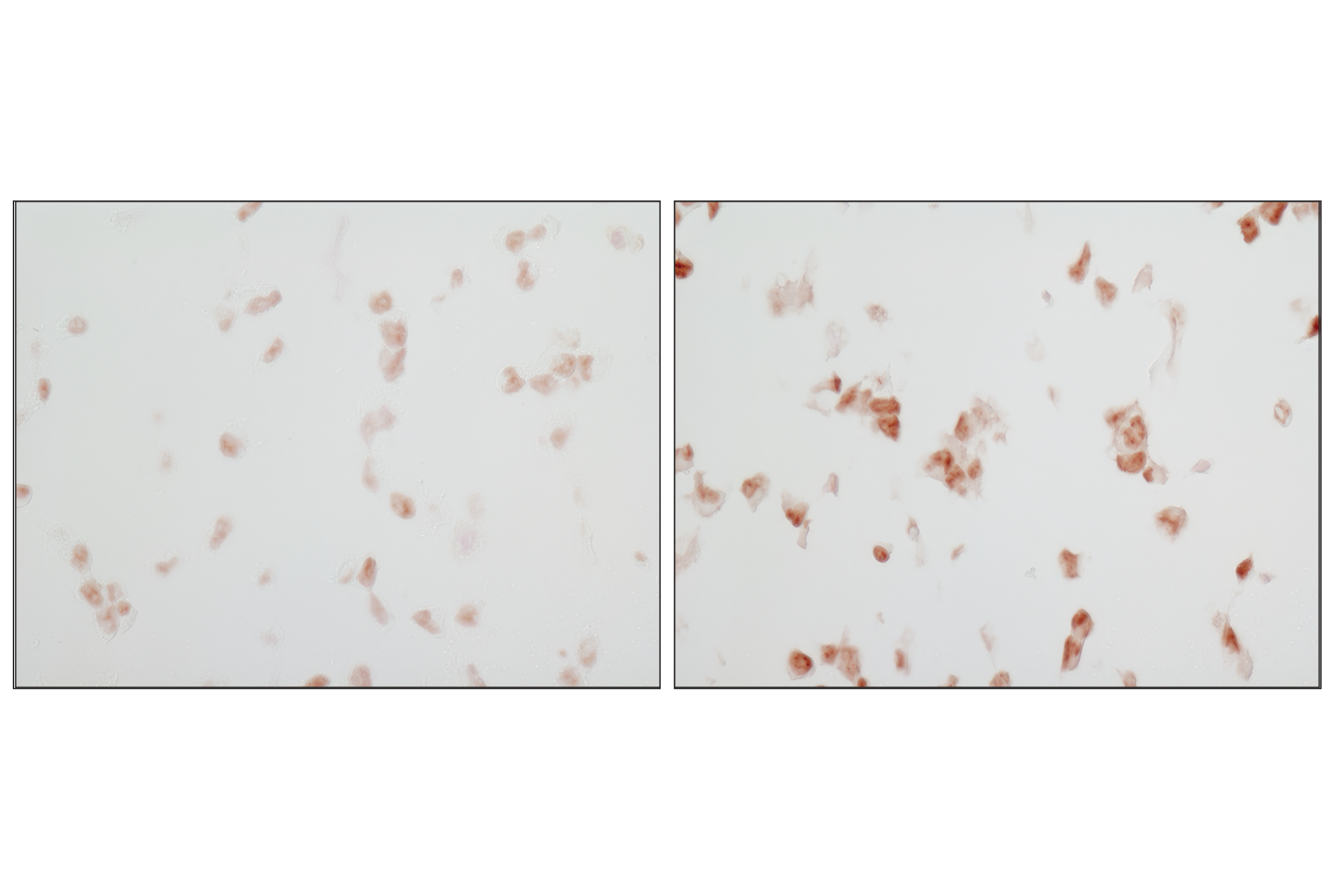
Immunohistochemical analysis of paraffin-embedded 293T cell pellets, untreated (left) or UV-treated (right), using Phospho-p38 MAPK (Thr180/Tyr182) (D3F9) XP® Rabbit mAb.
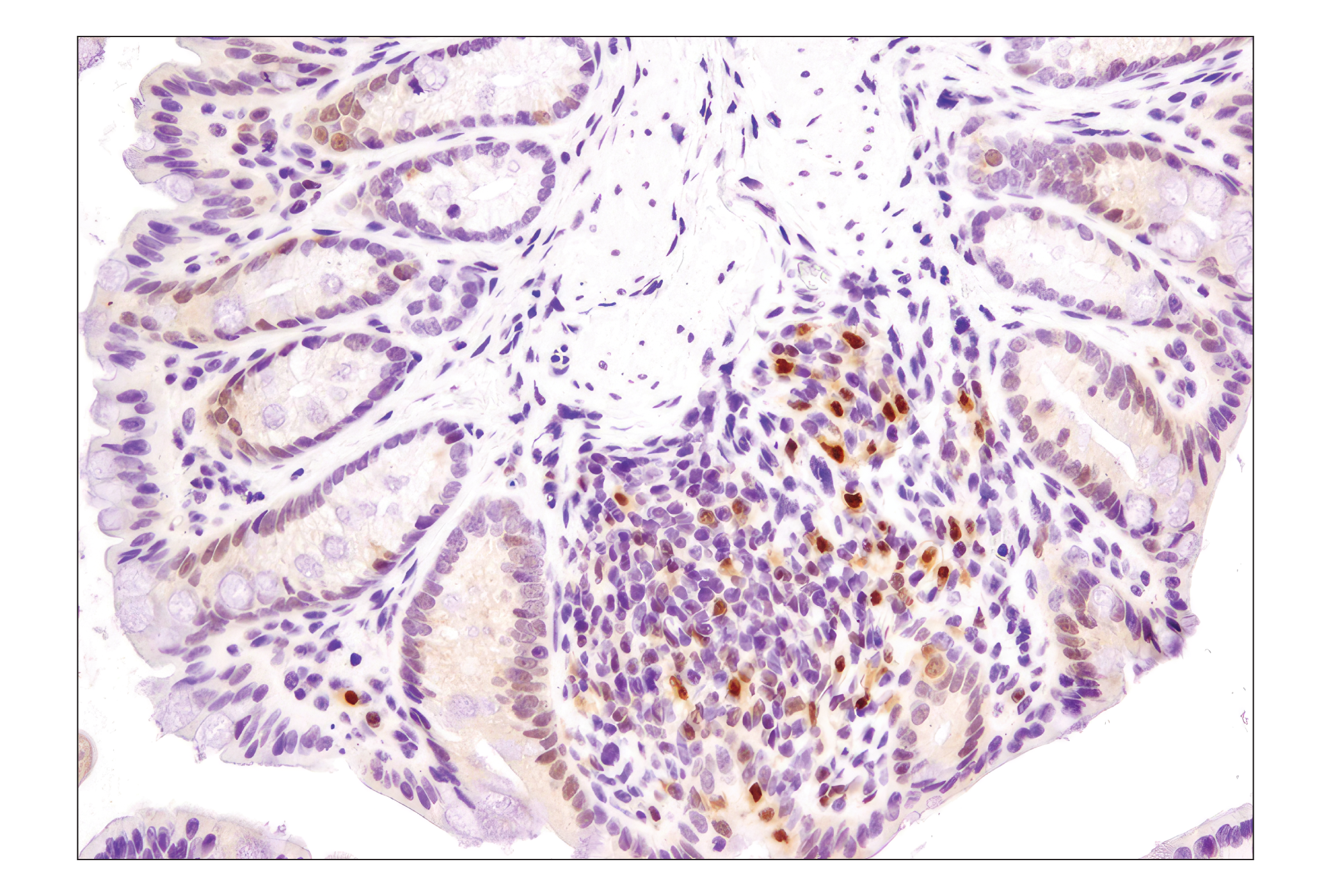
Immunohistochemical analysis of paraffin-embedded human colon carcinoma using Phospho-p38 MAPK (Thr180/Tyr182) (D3F9) XP® Rabbit mAb.
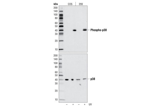
Western blot analysis of extracts from COS and 293 cells, untreated or UV-treated, using Phospho-p38 MAPK (Thr180/Tyr182) (D3F9) XP® Rabbit mAb (upper) or p38 MAPK Antibody #9212 (lower).
Western blot analysis of extracts from Xenopus oocytes overexpressing Myc-tagged p38 isoforms using p38α MAPK Antibody #9218 (upper) and Myc-Tag Antibody (lower) to demonstrate the isoform specificity of p38α MAPK Antibody. (Provided by Dr. Eusebio Perdiguero and Dr. Angel Nebreda, European Molecular Biology Laboratory, Germany.)
Western blot analysis of purified recombinant full-length p38 MAP kinase GST-fusion proteins using p38β MAP Kinase (C28C2) Rabbit mAb #2339 (upper), or a p38 MAP kinase pan antibody (lower).
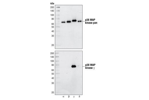
Western blot analysis of purified recombinant full-length p38 MAP kinase GST fusion proteins using p38 MAP kinase pan antibody (upper) or p38γ MAP Kinase Antibody #2307 (lower).
Western blot analysis of purified recombinant full-length p38 MAP kinase GST fusion proteins using p38 MAP kinase pan antibody (upper), or p38δ MAP Kinase (10A8) Rabbit mAb #2308 (lower).
Western blot analysis of extracts from COS and 293 cells, untreated or UV-treated, using Phospho-p38 MAPK (Thr180/Tyr182) (D3F9) XP® Rabbit mAb #4511 (upper) or p38 MAPK Antibody #9212 (lower).
Immunoprecipitation of phospho-p38 from THP1 cells, untreated or sorbitol-treated, using phospho-p38 MAPK (Thr180/Tyr182) Antibody #4511, followed by western blot with p38α Antibody #9218 (right two lanes). Left two lanes are THP1 whole cell extracts representing 10% of the IP input.
Immunoprecipitation of phospho-p38 from 293 cells, untreated, sorbitol or anisomycin-treated, using phospho-p38 MAPK (Thr180/Tyr182) Antibody #4511, followed by western blot with p38β Antibody #2339 (right three lanes). Left three lanes are 293 whole cell extracts representing 10% of the IP input. Western blot was detected using the Mouse Anti-rabbit IgG (Confirmation Specific) mAb #3678.
Immunoprecipitation of phospho-p38 from THP1 cells, untreated or sorbitol-treated, using phospho-p38 MAPK (Thr180/Tyr182) Antibody #4511, followed by western blot with p38γ Antibody #2307 (right two lanes). Left two lanes are THP1 whole cell extracts representing 10% of the IP input. Western blot was detected using the Mouse Anti-rabbit IgG (Confirmation Specific) mAb #3678.
Immunoprecipitation of phospho-p38 from 293 cells, untreated, sorbitol or anisomycin-treated, using phospho-p38 MAPK (Thr180/Tyr182) Antibody #4511, followed by western blot with p38δ Antibody #2308 (right three lanes). Left three lanes are 293 whole cell extracts representing 10% of the IP input. Western blot was detected using the Mouse Anti-rabbit IgG (Confirmation Specific) mAb #3678.
Immunohistochemical analysis of paraffin-embedded mouse colon using Phospho-p38 MAPK (Thr180/Tyr182) (D3F9) XP® Rabbit mAb.

 危险品化学品经营许可证(不带存储) 许可证编号:沪(杨)应急管危经许[2022]202944(QY)
  营业执照(三证合一)