


 全部商品分类
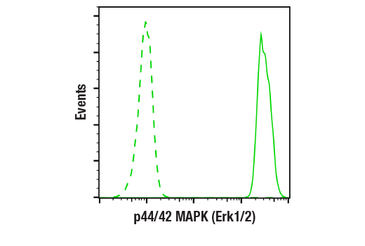


p44/42 MAPK (Erk1/2) (137F5) Rabbit mAb


 下载产品说明书 下载COA 下载SDS
 用小程序,查商品更便捷


 收藏
收藏
 对比
对比 咨询
咨询
Monoclonal antibody is produced by immunizing animals with a synthetic peptide corresponding to residues near the C-terminus of human p44 MAP kinase.
Product Usage Information
| Application | Dilution |
|---|---|
| Western Blotting | 1:1000 |
| Simple Western™ | 1:10 - 1:50 |
| Immunoprecipitation | 1:50 |
| Immunohistochemistry (Paraffin) | 1:125 - 1:500 |
| Immunofluorescence (Frozen) | 1:400 - 1:800 |
| Immunofluorescence (Immunocytochemistry) | 1:400 - 1:1600 |
| Flow Cytometry (Fixed/Permeabilized) | 1:200 - 1:800 |
Specificity/Sensitivity
Species Reactivity:
Human, Mouse, Rat, Hamster, Monkey, Mink, D. melanogaster, Zebrafish, Bovine, Dog, Pig, C. elegans
Supplied in 10 mM sodium HEPES (pH 7.5), 150 mM NaCl, 100 µg/ml BSA, 50% glycerol and less than 0.02% sodium azide. Store at –20°C. Do not aliquot the antibody.
For a carrier free (BSA and azide free) version of this product see product #68303.
参考图片
Flow cytometric analysis of Jurkat cells using p44/42 MAPK (Erk1/2) (137F5) Rabbit mAb (solid line) compared to concentration-matched Rabbit (DA1E) mAb IgG XP® Isotype Control #3900 (dashed line). Anti-rabbit IgG (H+L), F(ab')₂ Fragment (Alexa Fluor® 488 Conjugate) #4412 was used as a secondary antibody.
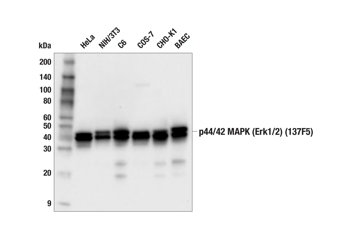
Western blot analysis of extracts from various cell lines using p44/42 MAPK (Erk1/2) (137F5) Rabbit mAb.
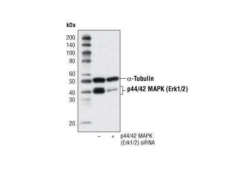
Western blot analysis of extracts from Hek 293 cells, transfected with 100 nM SignalSilence® Control siRNA (Fluorescein Conjugate) #6201 (-) or SignalSilence® p44/42 MAPK (Erk1/2) siRNA (+), using p44/42 MAPK (Erk1/2) (137F5) Rabbit mAb #4695 and α-Tubulin (11H10) Rabbit mAb #2125. The p44/42 MAPK (Erk1/2) (137F5) Rabbit mAb confirms silencing of p44/42 expression and α-Tubulin (11H10) Rabbit mAb is used to control for loading and specificity of p44/42 MAPK (Erk1/2) siRNA.
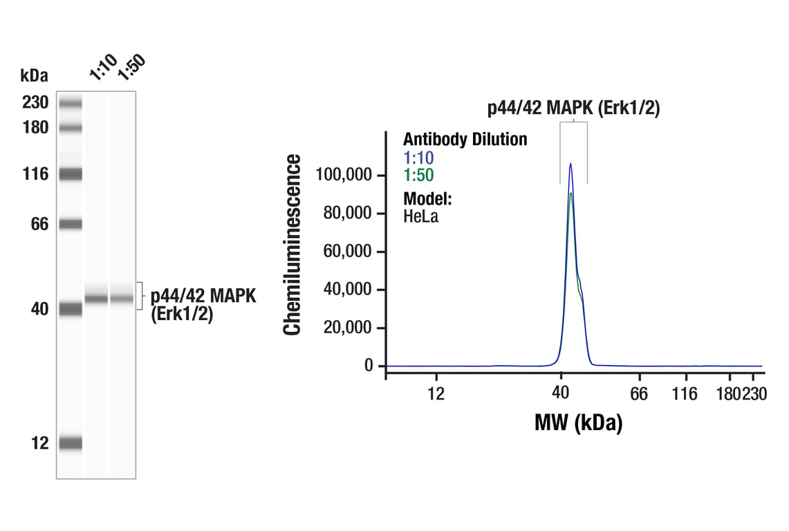
Simple Western™ analysis of lysates (0.1 mg/mL) from HeLa cells using p44/42 MAPK (Erk1/2) (137F5) Rabbit mAb #4695. The virtual lane view (left) shows the target band (as indicated) at 1:10 and 1:50 dilutions of primary antibody. The corresponding electropherogram view (right) plots chemiluminescence by molecular weight along the capillary at 1:10 (blue line) and 1:50 (green line) dilutions of primary antibody. This experiment was performed under reducing conditions on the Jess™ Simple Western instrument from ProteinSimple, a BioTechne brand, using the 12-230 kDa separation module.
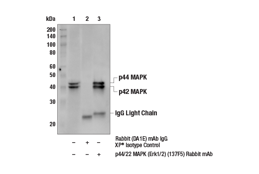
Immunoprecipitation of Jurkat cell extracts. Lane 1 is 10% input, lane 2 is Rabbit (DA1E) mAb IgG XP® Isotype Control #3900, and lane 3 is p44/42 MAPK (Erk1/2) (137F5) Rabbit mAb. Western blot analysis was performed using p44/42 MAPK (Erk1/2) (137F5) Rabbit mAb. Mouse Anti-Rabbit IgG (Light-Chain Specific) (D4W3E) mAb (HRP Conjugate) #93702 was used as a secondary antibody.
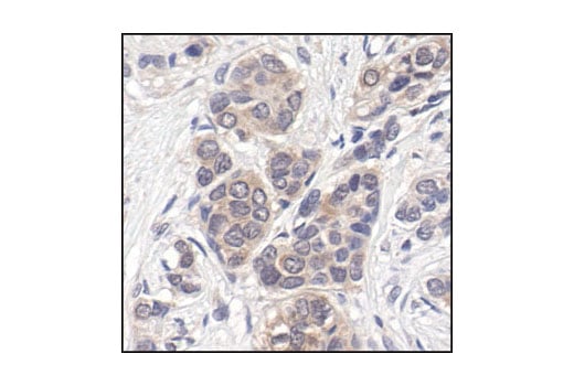
Immunohistochemical analysis of paraffin-embedded human breast carcinoma, showing cytoplasmic and nuclear localization, using p44/42 MAPK (Erk1/2) (137F5) Rabbit mAb.
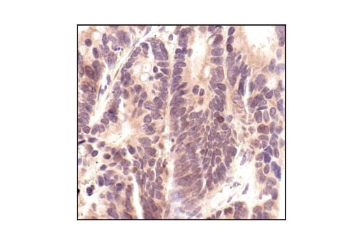
Immunohistochemical analysis of paraffin-embedded human colon carcinoma, using p44/42 MAPK (Erk1/2) (137F5) Rabbit mAb.
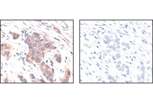
Immunohistochemical analysis of paraffin-embedded human breast carcinoma, using p44/42 MAPK (Erk1/2) (137F5) Rabbit mAb in the presence of control peptide (left) or #1240 p44/42 MAPK (Erk1/2) Blocking Peptide (#4695 Specific) (right).
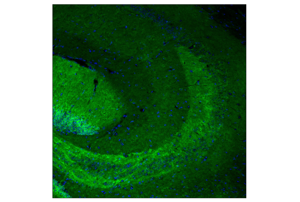
Confocal immunofluorescent analysis of fixed frozen mouse hippocampus using p44/42 MAPK (Erk1/2) (137F5) Rabbit mAb (green) and ProLong Gold Antifade Reagent with DAPI #8961 (blue).
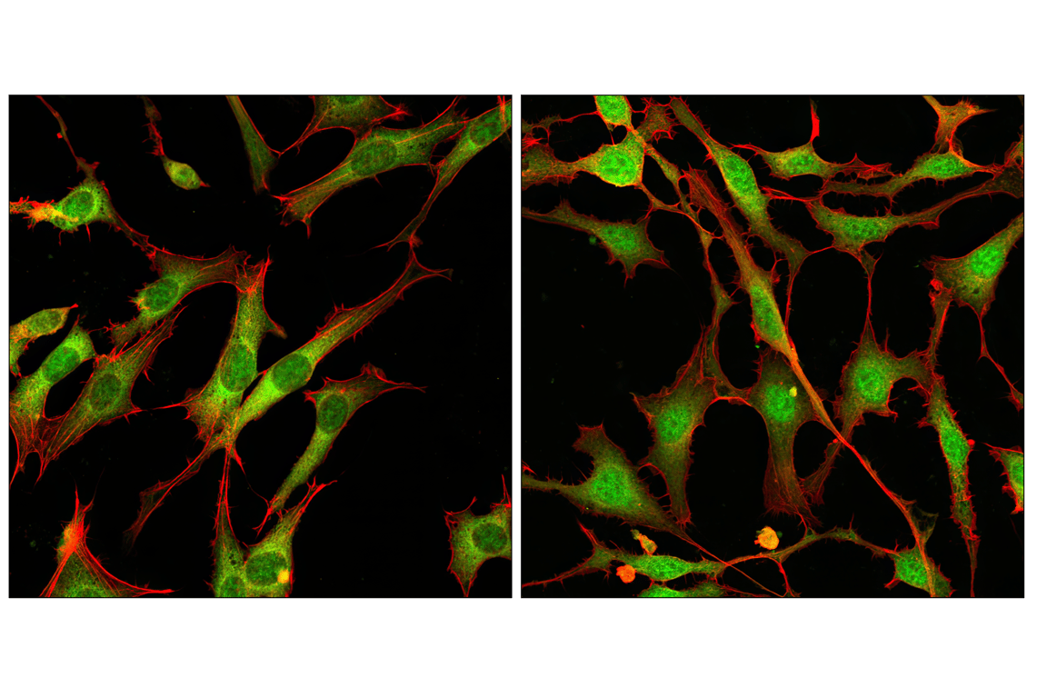
Confocal immunofluorescent analysis of NIH/3T3 cells, treated with either U0126 (MEK1/2 Inhibitor) #9903 (left) or PDGF (right), using p44/42 MAPK (Erk1/2) (137F5) Rabbit mAb (green). Actin filaments have been labeled with DY-554 phalloidin (red).