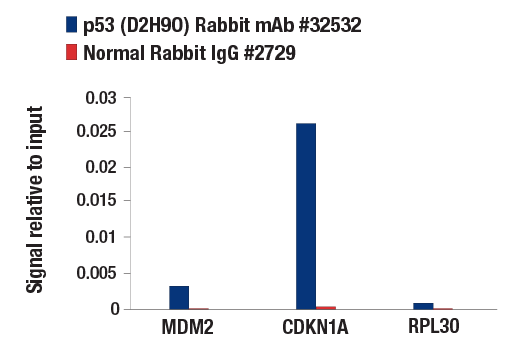
p53 (D2H9O) Rabbit mAb
1/3

p53 (D2H9O) Rabbit mAb

分享
品牌: CST
pdf 下载产品说明书
pdf 下载COA
pdf 下载SDS
用小程序,查商品更便捷
收藏
对比对比
咨询咨询
分子量:
53
来源宿主:
Rabbit
产品介绍
产品信息
荧光素标记
来源纯化

Monoclonal antibody is produced by immunizing animals with a synthetic peptide corresponding to residues surrounding Ala350 of mouse p53 protein.

宿主
Rabbit
商品描述

Product Usage Information

For optimal ChIP results, use 10 μl of antibody and 10 μg of chromatin (approximately 4 x 10⁶ cells) per IP. This antibody has been validated using SimpleChIP® Enzymatic Chromatin IP Kits.

ApplicationDilution
Western Blotting1:1000
Immunoprecipitation1:200
Chromatin IP1:50
同种型
Rabbit IgG
分子量
53
应用
目标/特异性

Specificity/Sensitivity

p53 (D2H9O) Rabbit mAb recognizes endogenous levels of total p53 protein.

Species Reactivity:

Mouse, Rat

敏感性
Endogenous
背景
背景
The p53 tumor suppressor protein plays a major role in cellular response to DNA damage and other genomic aberrations. Activation of p53 can lead to either cell cycle arrest and DNA repair or apoptosis (1). p53 is phosphorylated at multiple sites in vivo and by several different protein kinases in vitro (2,3). DNA damage induces phosphorylation of p53 at Ser15 and Ser20 and leads to a reduced interaction between p53 and its negative regulator, the oncoprotein MDM2 (4). MDM2 inhibits p53 accumulation by targeting it for ubiquitination and proteasomal degradation (5,6). p53 can be phosphorylated by ATM, ATR, and DNA-PK at Ser15 and Ser37. Phosphorylation impairs the ability of MDM2 to bind p53, promoting both the accumulation and activation of p53 in response to DNA damage (4,7). Chk2 and Chk1 can phosphorylate p53 at Ser20, enhancing its tetramerization, stability, and activity (8,9). p53 is phosphorylated at Ser392 in vivo (10,11) and by CAK in vitro (11). Phosphorylation of p53 at Ser392 is increased in human tumors (12) and has been reported to influence the growth suppressor function, DNA binding, and transcriptional activation of p53 (10,13,14). p53 is phosphorylated at Ser6 and Ser9 by CK1δ and CK1ε both in vitro and in vivo (13,15). Phosphorylation of p53 at Ser46 regulates the ability of p53 to induce apoptosis (16). Acetylation of p53 is mediated by p300 and CBP acetyltransferases. Inhibition of deacetylation suppressing MDM2 from recruiting HDAC1 complex by p19 (ARF) stabilizes p53. Acetylation appears to play a positive role in the accumulation of p53 protein in stress response (17). Following DNA damage, human p53 becomes acetylated at Lys382 (Lys379 in mouse) in vivo to enhance p53-DNA binding (18). Deacetylation of p53 occurs through interaction with the SIRT1 protein, a deacetylase that may be involved in cellular aging and the DNA damage response (19). 1.Levine, A.J. (1997) Cell 88, 323-31. 2.Meek, D.W. (1994) Semin Cancer Biol 5, 203-10. 3.Milczarek, G.J. et al. (1997) Life Sci 60, 1-11. 4.Shieh, S.Y. et al. (1997) Cell 91, 325-34. 5.Chehab, N.H. et al. (1999) Proc Natl Acad Sci U S A 96, 13777-82. 6.Honda, R. et al. (1997) FEBS Lett 420, 25-7. 7.Tibbetts, R.S. et al. (1999) Genes Dev 13, 152-7. 8.Shieh, S.Y. et al. (1999) EMBO J 18, 1815-23. 9.Hirao, A. et al. (2000) Science 287, 1824-7. 10.Hao, M. et al. (1996) J Biol Chem 271, 29380-5. 11.Lu, H. et al. (1997) Mol Cell Biol 17, 5923-34. 12.Ullrich, S.J. et al. (1993) Proc Natl Acad Sci U S A 90, 5954-8. 13.Kohn, K.W. (1999) Mol Biol Cell 10, 2703-34. 14.Lohrum, M. and Scheidtmann, K.H. (1996) Oncogene 13, 2527-39. 15.Knippschild, U. et al. (1997) Oncogene 15, 1727-36. 16.Oda, K. et al. (2000) Cell 102, 849-62. 17.Ito, A. et al. (2001) EMBO J 20, 1331-40. 18.Sakaguchi, K. et al. (1998) Genes Dev 12, 2831-41. 19.Solomon, J.M. et al. (2006) Mol Cell Biol 26, 28-38.
研究领域
癌症,细胞生物学,神经科学
翻译后修饰
unmodified
制备和贮存
保存方式

Supplied in 10 mM sodium HEPES (pH 7.5), 150 mM NaCl, 100 µg/ml BSA, 50% glycerol and less than 0.02% sodium azide. Store at –20°C. Do not aliquot the antibody.

数据库链接
Entrez-Gene ID
22059
UniProt ID
P02340

参考图片

Chromatin immunoprecipitations were performed with cross-linked chromatin from doxorubicin-treated (0.5 μM; 24 hr) 3T3 cells and either p53 (D2H9O) Rabbit mAb or Normal Rabbit IgG #2729, using SimpleChIP® Enzymatic Chromatin IP Kit (Magnetic Beads) #9003. The enriched DNA was quantified by real-time PCR using SimpleChIP® Mouse MDM2 Exon 3 Primers #76144, mouse CDKN1A promoter primers, and SimpleChIP® Mouse RPL30 Intron 2 Primers #7015. The amount of immunoprecipitated DNA in each sample is represented as signal relative to the total amount of input chromatin (equivalent to one).

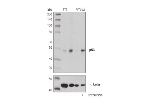
Western blot analysis of extracts from 3T3 and MTLN3 cells, untreated (-) or treated with doxorubicin (0.5 μM; 24 hr; +), using p53 (D2H9O) Rabbit mAb (upper) or β-Actin (D6A8) Rabbit mAb #8457 (lower).

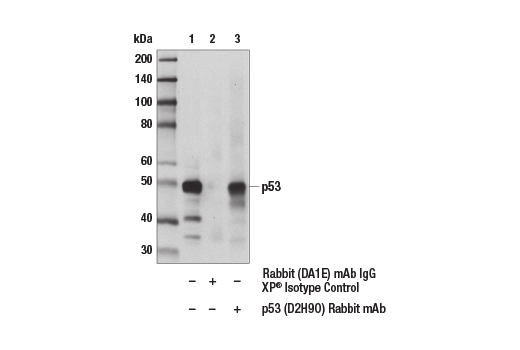
Immuonprecipitation of p53 from doxorubicin-treated (0.5 μM; 24 hr) 3T3 cell extracts. Lane 1 is 10% input, lane 2 is Rabbit (DA1E) mAb IgG XP® Isotype Control #3900, and lane 3 is p53 (D2H9O) Rabbit mAb. Western blot analysis was performed using p53 (D2H9O) Rabbit mAb. A light chain-specific secondary antibody was used.

声明 :本官网所有报价均为常温或者蓝冰运输价格,如有产品需要干冰运输,需另外加收干冰运输费。
货号:
32532T
一键复制
询价
供应商现货
20μl
100ul
选择数量
当前规格1件起购 
配送至
预计5-6个工作日送达,快递: 免运费,若需干冰额外收费