


 全部商品分类


 用小程序,查商品更便捷


Monoclonal antibody is produced by immunizing animals with a full-length human p53 fusion protein.
Product Usage Information
For optimal ChIP results, use 2.5 μl of antibody and 10 μg of chromatin (approximately 4 x 10⁶ cells) per IP. This antibody has been validated using SimpleChIP® Enzymatic Chromatin IP Kits.
| Application | Dilution |
|---|---|
| Western Blotting | 1:1000 |
| Simple Western™ | 1:10 - 1:50 |
| Immunohistochemistry (Paraffin) | 1:80 - 1:320 |
| Immunofluorescence (Immunocytochemistry) | 1:800 - 1:1600 |
| Flow Cytometry (Fixed/Permeabilized) | 1:800 - 1:3200 |
| Chromatin IP | 1:200 |
Specificity/Sensitivity
Species Reactivity:
Human, Monkey
Supplied in 10 mM sodium HEPES (pH 7.5), 150 mM NaCl, 100 µg/ml BSA, 50% glycerol and less than 0.02% sodium azide. Store at –20°C. Do not aliquot the antibody.
For a carrier free (BSA and azide free) version of this product see product #74556
参考图片
Chromatin immunoprecipitations were performed with cross-linked chromatin from HCT116 cells treated with UV (100 J/m2 followed by a 3 hour recovery) and either p53 (7F5) Rabbit mAb or Normal Rabbit IgG #2729 using SimpleChIP® Enzymatic Chromatin IP Kit (Magnetic Beads) #9003. The enriched DNA was quantified by real-time PCR using SimpleChIP® Human CDKN1A Promoter Primers #6449, human MDM2 intron 2 primers, and SimpleChIP® Human α Satellite Repeat Primers #4486. The amount of immunoprecipitated DNA in each sample is represented as signal relative to the total amount of input chromatin, which is equivalent to one.
Western blot analysis of extracts from 293 and COS cells, using p53 (7F5) Rabbit mAb.
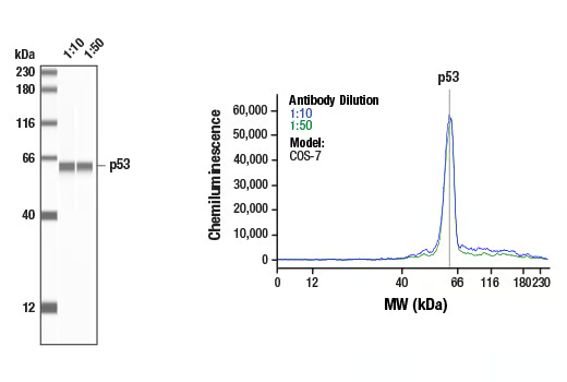
Simple Western™ analysis of lysates (1 mg/mL) from COS-7 cells using p53 (7F5) Rabbit mAb #2527. The virtual lane view (left) shows the target band (as indicated) at 1:10 and 1:50 dilutions of primary antibody. The corresponding electropherogram view (right) plots chemiluminescence by molecular weight along the capillary at 1:10 (blue line) and 1:50 (green line) dilutions of primary antibody. This experiment was performed under reducing conditions on the Jess™ Simple Western instrument from ProteinSimple, a BioTechne brand, using the 12-230 kDa separation module.
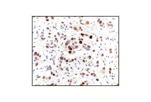
Immunohistochemical analysis of paraffin-embedded human breast carcinoma, using p53 (7F5) Rabbit mAb.
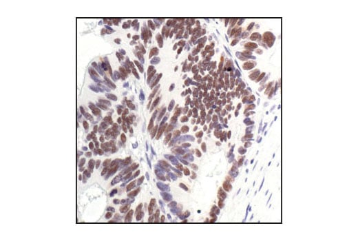
Immunohistochemical analysis of paraffin-embedded human colon carcinoma, using p53 (7F5) Rabbit mAb.
Immunohistochemical analysis of paraffin-embedded HT-29 (left) and SaOs-2 (right) cells, using p53 (7F5) Rabbit mAb. Note the lack of staining in p53-negative SaOs-2 cells.
Confocal Immunofluorescent analysis of HT-29 cells using p53 (7F5) Rabbit mAb (green). Actin filaments have been labeled with DY-554 phalloidin (red).
Flow cytometric analysis of HT-29 cells using p53 (7F5) Rabbit mAb (solid line) compared to concentration-matched Rabbit (DA1E) mAb IgG XP® Isotype Control #3900 (dashed line). Anti-rabbit IgG (H+L), F(ab')2 Fragment (Alexa Fluor® 488 Conjugate) #4412 was used as a secondary antibody.