


 全部商品分类


p53 Antibody Sampler Kit

 下载产品说明书 下载COA
 用小程序,查商品更便捷


 收藏
收藏
 对比
对比 咨询
咨询
The p53 Antibody Sampler Kit provides an economical means of detecting p53 activity using modification-specific and control antibodies. The kit includes enough antibody to perform two western blot experiments with each primary antibody.
Specificity/Sensitivity
参考图片
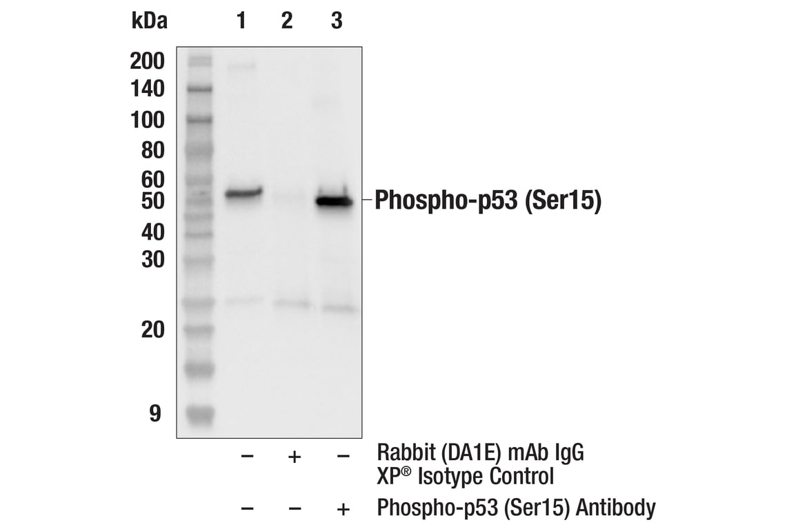
Immunoprecipitation of Phospho-p53 (Ser15) protein from HT-29 + Etoposide #2200 (25µM, Overnight) cell extracts. Lane 1 is 10% input, lane 2 is Normal Rabbit IgG #2729, and lane 3 is Phospho-p53 (Ser15) Antibody. Western blot analysis was performed using Phospho-p53 (Ser15) Antibody. Mouse Anti-rabbit IgG (Conformation Specific) (L27A9) mAb #3678 was used as a secondary antibody. Note that Phospho-p53 (Ser15) Antibody cross-reacts with some proteins present in the Blue Prestained Protein Marker, Broad Range (11-250 kDa) #59329. In this figure, only proteins present in the Biotinylated Protein Ladder Detection Pack #7727 are labeled.
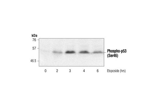
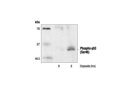
Western blot analysis of extracts from MCF-7 cells treated with etoposide for the indicated times, using Phospho-p53 (Ser46) Antibody.
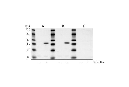
Western blot analysis of extracts from HeLa cells, untreated or treated with both trichostatin A #9950 (400 nM for 24 hours), and doxorubicin (0.5 µM for 24 hours) using Acetyl-p53 (Lys382) Antibody alone (A), antibody preincubated with a non-acetylated Lys382 peptide (B), or antibody preincubated with an acetylated Lys382 peptide (C).
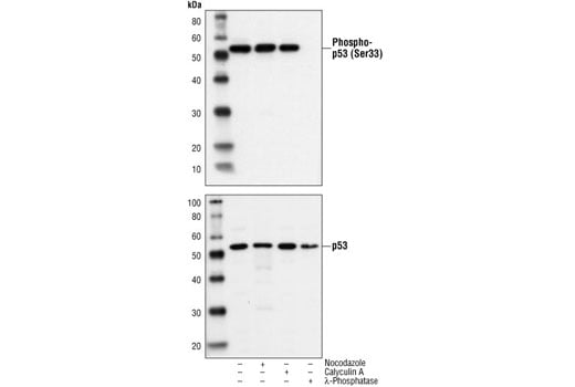
Western blot analysis of extracts from HT29 cells treated with nocodazole (50 ng/ml, 24h), calyculin A #9902 (100 nM, 10 min), or lambda Phosphatase NEB#P0753 (10,000 units/ml, 1 h), using Phospho-p53 (Ser33) Antibody (upper) or p53 (1C12) Mouse mAb #2524 (lower).
Western blot analysis of extracts from 293 and COS cells, using p53 (7F5) Rabbit mAb.
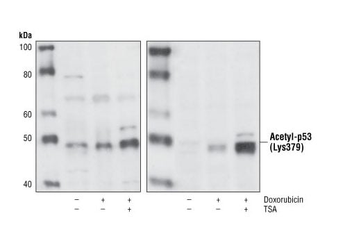
Western blot analysis of extracts from NIH/3T3 (left) and MCF-7 (right) cells, untreated, doxorubicin-treated (0.5 μM, 24 hours) or doxorubicin and trichostatin A-treated (TSA, #9950, 400 nM, 24 hours), using Acetyl-p53 (Lys379) Antibody.
After the primary antibody is bound to the target protein, a complex with HRP-linked secondary antibody is formed. The LumiGLO® is added and emits light during enzyme catalyzed decomposition.
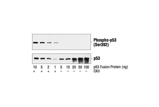
Western blot analysis of increasing amounts of a p53 fusion protein, untreated or phosphorylated by CKII, using Phospho-p53 (Ser392) Antibody (upper) or p53 Antibody #9282 (lower).
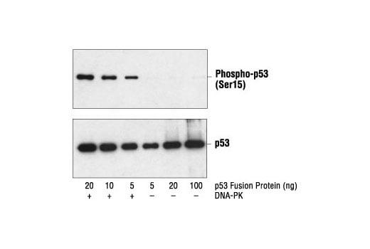
Western blot analysis of a p53 fusion protein, untreated or phosphorylated by DNA-PK, using Phospho-p53 (Ser15) Antibody (upper) and p53 Antibody #9282 (lower).
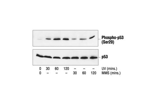
Western blot analysis of extracts from COS cells treated with UV or MMS for the indicated times, using Phospho-p53 (Ser20) Antibody (upper) or p53 Antibody #9282 (lower).
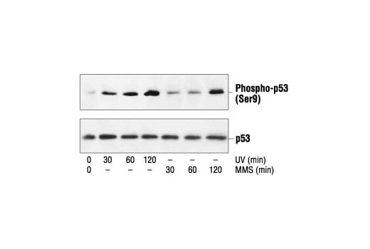
Western blot analysis of extracts from COS cells treated with UV or MMS for the indicated times, using Phospho-p53 (Ser9) Antibody (upper) or p53 Antibody #9282 (lower).
Immunoprecipitation of extracts from MCF-7 cells treated with etoposide under nondenaturing conditions, using Phospho-p53 (Ser46) Antibody, followed by Western blot analysis using a monoclonal p53 antibody.
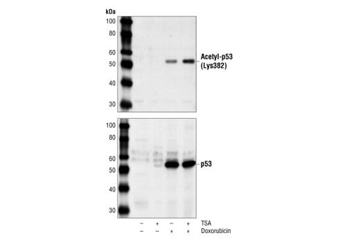
Western blot analysis of extracts from HeLa cells, untreated, trichostatin A-treated #9950 (400 nM for 24 hours), doxorubicin-treated (0.5 µM for 24 hours), or both, using Acetyl-p53 (Lys382) Antibody (top) or p53 Antibody #2524 (bottom).
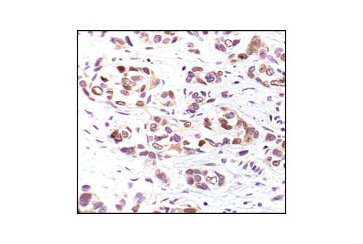
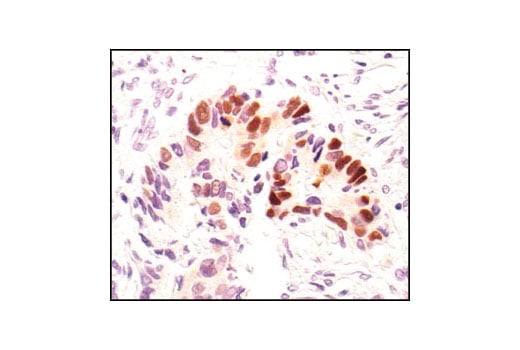
Immunohistochemical analysis of paraffin-embedded human breast carcinoma, using Phospho-p53 (Ser33) Antibody.
Immunohistochemical analysis of paraffin-embedded human breast carcinoma, using p53 (7F5) Rabbit mAb.
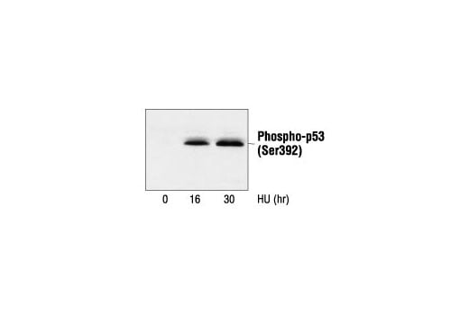
Western blot analysis of extracts from Mv1Lu cells, untreated or hydroxyurea-treated (20 mM), using Phospho-p53 (Ser392) Antibody.
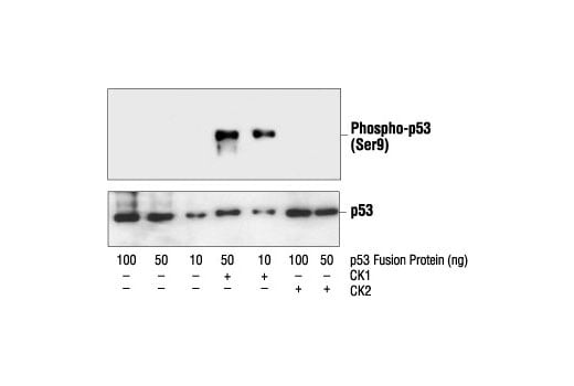
Western blot analysis of a p53 fusion protein, untreated or phosphorylated by CK1 or CK2, using Phospho-p53 (Ser9) Antibody (upper) or p53 Antibody #9282 (lower).
Confocal immunofluorescent analysis of MCF-7 cells, untreated (left) or etoposide-treated (right), using Phospho-p53 (Ser46) Antibody (green). Actin filaments have been labeled with DY-554 phalloidin (red).
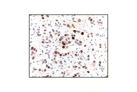
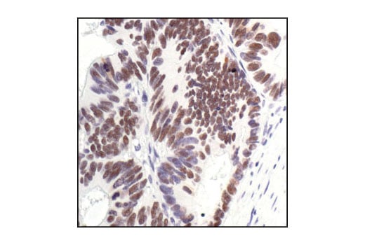
Immunohistochemical analysis of paraffin-embedded human colon carcinoma, showing nuclear localization, using Phospho-p53 (Ser33) Antibody.
Immunohistochemical analysis of paraffin-embedded human colon carcinoma, using p53 (7F5) Rabbit mAb.
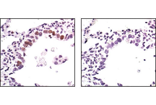
Immunohistochemical analysis of paraffin-embedded human lung dysplasia of alveolar cells, using Phospho-p53 (Ser33) Antibody in the presence of control peptide (left) or antigen specific peptide (right),
Immunohistochemical analysis of paraffin-embedded HT-29 (left) and SaOs-2 (right) cells, using p53 (7F5) Rabbit mAb. Note the lack of staining in p53-negative SaOs-2 cells.
Confocal Immunofluorescent analysis of HT-29 cells using p53 (7F5) Rabbit mAb (green). Actin filaments have been labeled with DY-554 phalloidin (red).
Flow cytometric analysis of HT-29 cells using p53 (7F5) Rabbit mAb (solid line) compared to concentration-matched Rabbit (DA1E) mAb IgG XP® Isotype Control #3900 (dashed line). Anti-rabbit IgG (H+L), F(ab')2 Fragment (Alexa Fluor® 488 Conjugate) #4412 was used as a secondary antibody.
Chromatin immunoprecipitations were performed with cross-linked chromatin from HCT116 cells treated with UV (100 J/m2 followed by a 3 hour recovery) and either p53 (7F5) Rabbit mAb or Normal Rabbit IgG #2729 using SimpleChIP® Enzymatic Chromatin IP Kit (Magnetic Beads) #9003. The enriched DNA was quantified by real-time PCR using SimpleChIP® Human CDKN1A Promoter Primers #6449, human MDM2 intron 2 primers, and SimpleChIP® Human α Satellite Repeat Primers #4486. The amount of immunoprecipitated DNA in each sample is represented as signal relative to the total amount of input chromatin, which is equivalent to one.
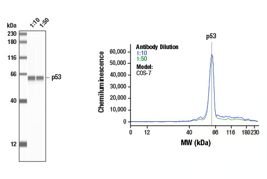
Simple Western™ analysis of lysates (1 mg/mL) from COS-7 cells using p53 (7F5) Rabbit mAb #2527. The virtual lane view (left) shows the target band (as indicated) at 1:10 and 1:50 dilutions of primary antibody. The corresponding electropherogram view (right) plots chemiluminescence by molecular weight along the capillary at 1:10 (blue line) and 1:50 (green line) dilutions of primary antibody. This experiment was performed under reducing conditions on the Jess™ Simple Western instrument from ProteinSimple, a BioTechne brand, using the 12-230 kDa separation module.
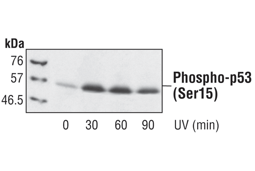
Western blot analysis of extracts from PC12 cells treated with UV for the indicated times, using Phospho-p53 (Ser15) Antibody.
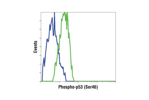
Flow cytometric analysis of HT-29 cells, untreated (blue) or UV-treated (green), using Phospho-p53 (Ser46) Antibody.
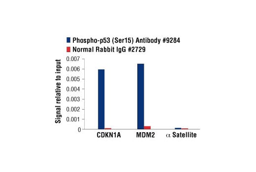
Chromatin immunoprecipitations were performed with cross-linked chromatin from HCT116 cells treated with UV (100 J/m2 followed by a 3 hour recovery) and either Phospho-p53 (Ser15) Antibody or Normal Rabbit IgG #2729 using SimpleChIP® Enzymatic Chromatin IP Kit (Magnetic Beads) #9003. The enriched DNA was quantified by real-time PCR using SimpleChIP® Human CDKN1A Promoter Primers #6449, human MDM2 intron 2 primers, and SimpleChIP® Human α Satellite Repeat Primers #4486. The amount of immunoprecipitated DNA in each sample is represented as signal relative to the total amount of input chromatin, which is equivalent to one.