

 全部商品分类
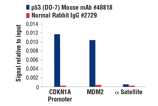


 用小程序,查商品更便捷


Monoclonal antibody is produced by immunizing animals with recombinant human p53 protein expressed in E. coli.

Product Usage Information
For optimal ChIP results, use 5 μl of antibody and 10 μg of chromatin (approximately 4 x 10⁶ cells) per IP. This antibody has been validated using SimpleChIP® Enzymatic Chromatin IP Kits.
| Application | Dilution |
|---|---|
| Western Blotting | 1:1000 |
| Immunohistochemistry (Paraffin) | 1:100 |
| Immunofluorescence (Immunocytochemistry) | 1:400 |
| Flow Cytometry (Fixed/Permeabilized) | 1:50 |
| Chromatin IP | 1:100 |
Specificity/Sensitivity
Species Reactivity:
Human
Supplied in 10 mM sodium HEPES (pH 7.5), 150 mM NaCl, 100 µg/ml BSA, 50% glycerol and less than 0.02% sodium azide. Store at –20°C. Do not aliquot the antibody.
For a carrier-free (BSA and azide free) version of this product see product #46565.
参考图片
Chromatin immunoprecipitations were performed with cross-linked chromatin from HCT 116 cells treated with UV (100 J/m2 followed by a 3 hr recovery) and either p53 (DO-7) Mouse mAb or Normal Rabbit IgG #2729 using SimpleChIP® Enzymatic Chromatin IP Kit (Magnetic Beads) #9003. The enriched DNA was quantified by real-time PCR using SimpleChIP® Human CDKN1A Promoter Primers #6449, human MDM2 intron 2 primers, and SimpleChIP® Human α Satellite Repeat Primers #4486. The amount of immunoprecipitated DNA in each sample is represented as signal relative to the total amount of input chromatin, which is equivalent to one.
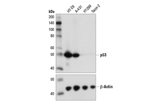
Western blot analysis of extracts from various cell lines using p53 (DO-7) Mouse mAb (upper) and β-Actin (D6A8) Rabbit mAb #8457 (lower).
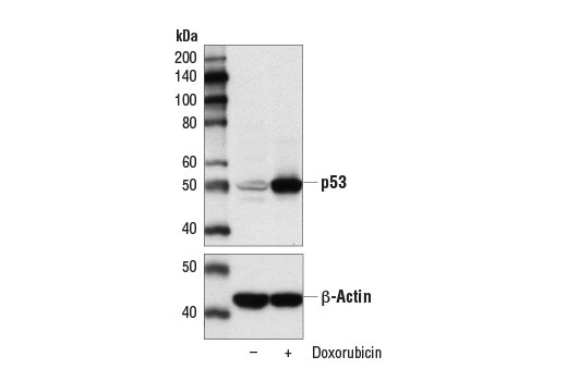
Western blot analysis of extracts from MCF7 cells, untreated (-) or treated with Doxorubicin #5927 (0.5 μM, 24 hr; +), using p53 (DO-7) Mouse mAb (upper) and β-Actin (D6A8) Rabbit mAb #8457 (lower).
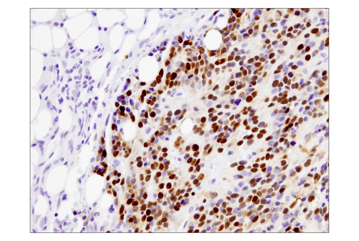
Immunohistochemical analysis of paraffin-embedded human breast carcinoma using p53 (DO-7) Mouse mAb.
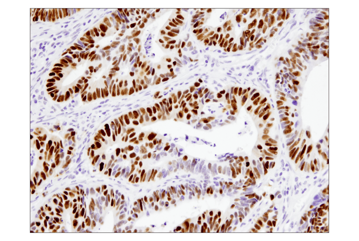
Immunohistochemical analysis of paraffin-embedded human colon carcinoma using p53 (DO-7) Mouse mAb.
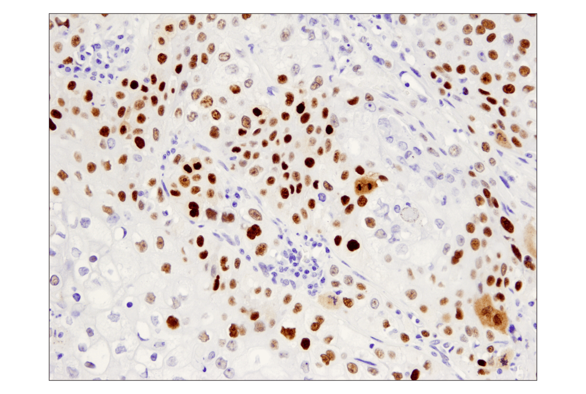
Immunohistochemical analysis of paraffin-embedded human squamous cell lung carcinoma using p53 (DO-7) Mouse mAb.
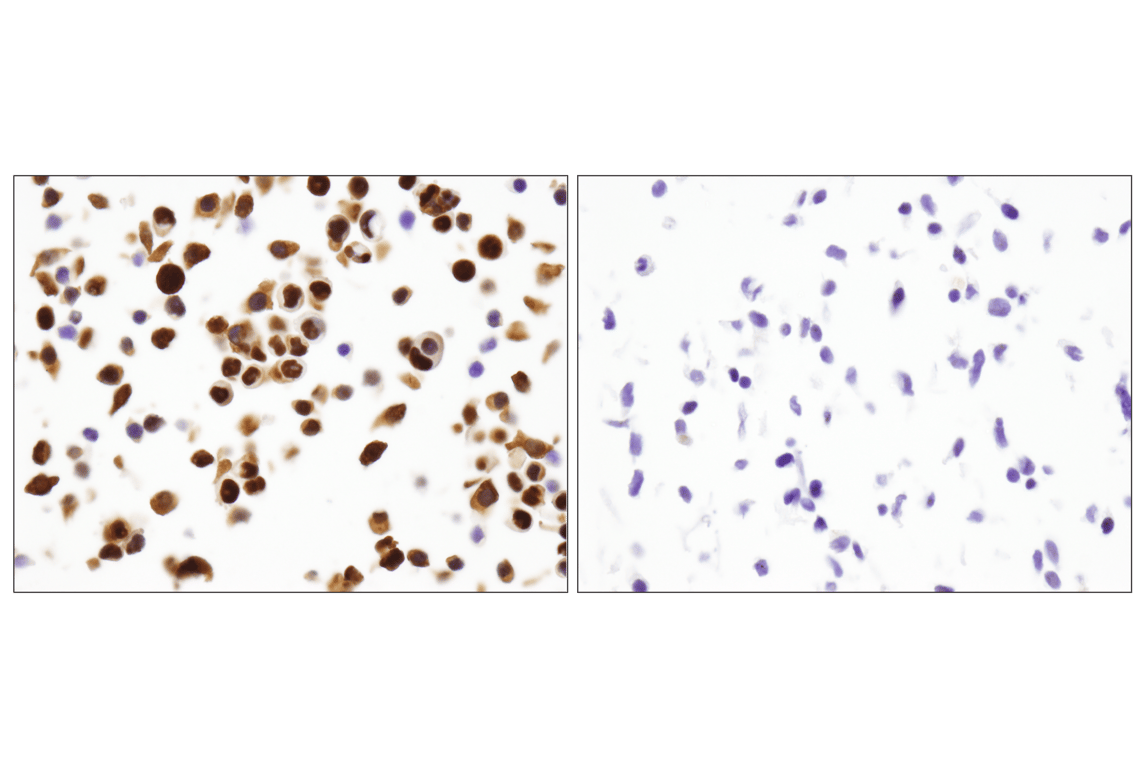
Immunohistochemical analysis of paraffin-embedded HT-29 (left) and Saos-2 (right) cells using p53 (DO-7) Mouse mAb.
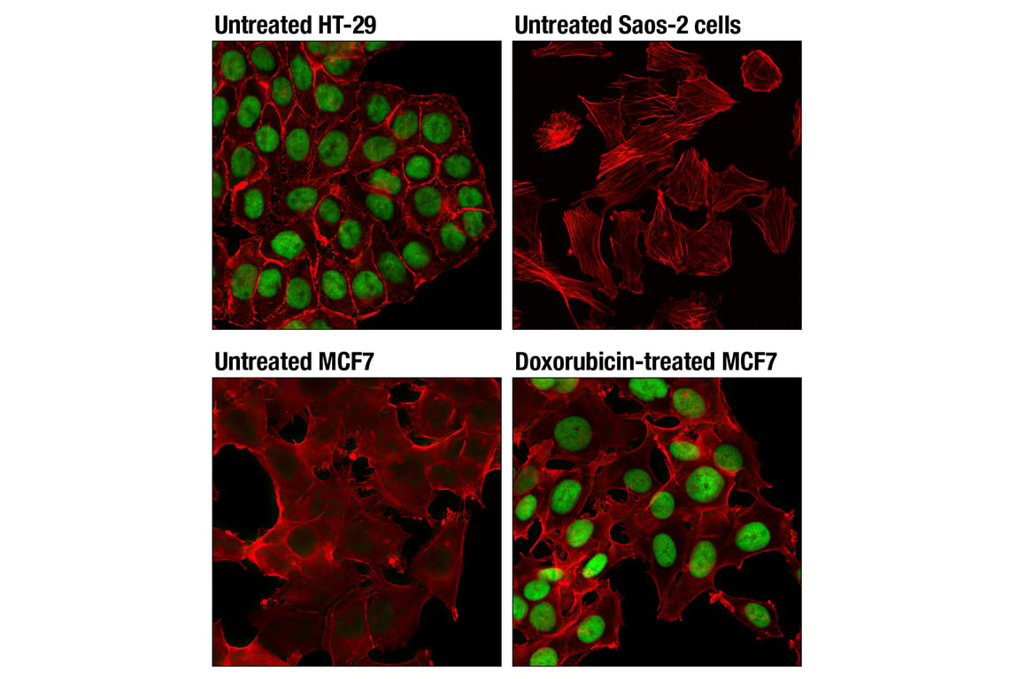
Confocal immunofluorescent analysis of untreated HT-29 cells (top left), untreated Saos-2 cells (top right), untreated MCF7 cells (bottom left), and MCF7 cells treated with Doxorubicin #5927 (0.5 μM, 24 hr; bottom right), using p53 (DO-7) Mouse mAb (green). Actin filaments were labeled with DyLight™ 554 Phalloidin #13054 (red).
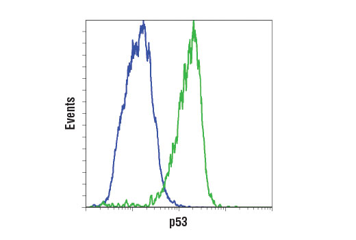
Flow cytometric analysis of H1299 cells (blue) and HT-29 cells (green) using p53 (DO-7) Mouse mAb. Anti-mouse IgG (H+L), F(ab')2 Fragment (Alexa Fluor® 488 Conjugate) #4408 was used as a secondary antibody.