PARP [Poly(ADP-Ribose) Polymerase] is a 113 kDa nuclear chromatin associated enzyme that catalyzes the transfer of ADP-ribose units from NAD+ to a variety of nuclear proteins including topoisomerases, histones, and PARP itself. The catalytic activity of PARP is increased in nonapoptotic cells following DNA damage, and PARP is thought to play an important role in mediating the normal cellular reponse to DNA damage and is a target of the caspase protease activity associated with apoptosis. During apoptosis, PARP is cleaved from a 113 kDa intact form into 89 kDa and 24 kDa fragments. This process separates the amino-terminal DNA-binding domain of the enzyme from the C-terminal catalytic domain resulting in the loss of normal PARP function. Although the role of PARP in apoptosis remains to be elucidated, PARP cleavage is considered to be a marker of apoptosis. This antibody has been reported to recognize an epitope located within the DNA-binding domain of the enzyme.
商品描述
C2-10
PARP [Poly(ADP-Ribose) Polymerase] is a 113 kDa nuclear chromatin associated enzyme that catalyzes the transfer of ADP-ribose units from NAD+ to a variety of nuclear proteins including topoisomerases, histones, and PARP itself. The catalytic activity of PARP is increased in nonapoptotic cells following DNA damage, and PARP is thought to play an important role in mediating the normal cellular reponse to DNA damage and is a target of the caspase protease activity associated with apoptosis. During apoptosis, PARP is cleaved from a 113 kDa intact form into 89 kDa and 24 kDa fragments. This process separates the amino-terminal DNA-binding domain of the enzyme from the C-terminal catalytic domain resulting in the loss of normal PARP function. Although the role of PARP in apoptosis remains to be elucidated, PARP cleavage is considered to be a marker of apoptosis. This antibody has been reported to recognize an epitope located within the DNA-binding domain of the enzyme.
同种型
Mouse IgG1
克隆号
克隆 C2-10 (RUO)
应用
实验应用
Western blot (Routinely Tested), Immunofluorescence (Reported)
研发参考(5)
1. Kaufmann SH, Desnoyers S, Ottaviano Y, Davidson NE, Poirier GG. Specific proteolytic cleavage of poly(ADP-ribose) polymerase: an early marker of chemotherapy-induced apoptosis. Cancer Res. 1993; 53(17):3976-3985. (Biology: Western blot).
2. Lamarre D, Talbot B, Leduc Y, Muller S, Poirier G. Production and characterization of monoclonal antibodies specific for the functional domains of poly(ADP-ribose) polymerase. Biochem Cell Biol. 1986; 64(4):368-376. (Immunogen).
3. Lamarre D, Talbot B, de Murcia G, et al. Structural and functional analysis of poly(ADP ribose) polymerase: an immunological study. Biochim Biophys Acta. 1988; 950(2):147-160. (Clone-specific: Immunofluorescence).
4. Patel T, Gores GJ, Kaufmann SH. The role of proteases during apoptosis. FASEB J. 1996; 10(5):587-597. (Biology).
5. Tewari M, Quan LT, O'Rourke K, et al. Yama/CPP32 beta, a mammalian homolog of CED-3, is a CrmA-inhibitable protease that cleaves the death substrate poly(ADP-ribose) polymerase. Cell. 1995; 81(5):801-809. (Biology: Western blot).
参考图片
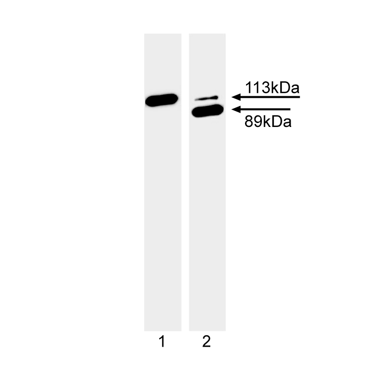
Western blot analysis of PARP cleavage. Jurkat cells (Human T-cell leukemia; ATCC TIB-152) were induced to undergo CD95 (Fas) mediated apoptosis by treatment with mouse anti-human CD95 antibody (clone DX2) (MN 555670) and Protein G for 4 hr (lane 2) or were untreated (lane 1). Lysates were probed with the purified mouse anti-PARP antibody (clone C2-10) at a dilution of 1:2000. The 116 kDa intact form of PARP is observed in both the untreated and anti-CD95 treated cell lysates. However, the 89 kDa PARP cleavage fragment is seen only in the treated cell lysates.
Western blot analysis of PARP cleavage. Jurkat cells (Human T-cell leukemia; ATCC TIB-152) were induced to undergo CD95 (Fas) mediated apoptosis by treatment with mouse anti-human CD95 antibody (clone DX2) (MN 555670) and Protein G for 4 hr (lane 2) or were untreated (lane 1). Lysates were probed with the purified mouse anti-PARP antibody (clone C2-10) at a dilution of 1:2000. The 116 kDa intact form of PARP is observed in both the untreated and anti-CD95 treated cell lysates. However, the 89 kDa PARP cleavage fragment is seen only in the treated cell lysates.


 全部商品分类


 下载产品说明书
 用小程序,查商品更便捷


 收藏
收藏
 对比
对比 咨询
咨询