PARP (Poly [ADP-Ribose] Polymerase) is a 113-kDa nuclear chromatin-associated enzyme that catalyzes the transfer of ADP-ribose units from NAD
+
to a variety of nuclear proteins including topoisomerases, histones, and PARP itself. The catalytic activity of PARP is increased in cells following DNA damage, and PARP is thought to play an important role in mediating the normal cellular response to DNA damage. Additionally, PARP is a target of the caspase protease activity associated with apoptosis. The PARP protein consists of an N-terminal DNA-binding domain (DBD) and a C-terminal catalytic domain separated by a central automodification domain. During apoptosis, Caspase-3 cleaves PARP at a recognition site (Asp Glu Val Asp Gly) in the DBD to form 24- and 89-kDa fragments. This process separates the DBD (which is mostly in the 24-kDa fragment) from the catalytic domain (in the 89-kDa fragment) of the enzyme, resulting in the loss of normal PARP function. It has been proposed that inactivation of PARP directs DNA-damaged cells to undergo apoptosis rather than necrotic degradation, and the presence of the 89-kDa PARP cleavage fraction is considered to be a marker of apoptosis.
A peptide corresponding to the N-terminus of the cleavage site (Asp 214) of human PARP was used as the immunogen. The F21-852 monoclonal antibody reacts only with the 89-kDa fragment of human PARP-1 that is downstream of the Caspase-3 cleavage site (Asp214) and contains the automodification and catalytic domains. It does not react with intact human PARP-1. Cross-reactivity with other members of the PARP superfamily is unknown. Recognition of cleaved PARP in mouse cells has been demonstrated, and it may also cross-react with a number of other species due to the conserved nature of the molecule.
商品描述
F21-852
PARP (Poly [ADP-Ribose] Polymerase) is a 113-kDa nuclear chromatin-associated enzyme that catalyzes the transfer of ADP-ribose units from NAD
+
to a variety of nuclear proteins including topoisomerases, histones, and PARP itself. The catalytic activity of PARP is increased in cells following DNA damage, and PARP is thought to play an important role in mediating the normal cellular response to DNA damage. Additionally, PARP is a target of the caspase protease activity associated with apoptosis. The PARP protein consists of an N-terminal DNA-binding domain (DBD) and a C-terminal catalytic domain separated by a central automodification domain. During apoptosis, Caspase-3 cleaves PARP at a recognition site (Asp Glu Val Asp Gly) in the DBD to form 24- and 89-kDa fragments. This process separates the DBD (which is mostly in the 24-kDa fragment) from the catalytic domain (in the 89-kDa fragment) of the enzyme, resulting in the loss of normal PARP function. It has been proposed that inactivation of PARP directs DNA-damaged cells to undergo apoptosis rather than necrotic degradation, and the presence of the 89-kDa PARP cleavage fraction is considered to be a marker of apoptosis.
A peptide corresponding to the N-terminus of the cleavage site (Asp 214) of human PARP was used as the immunogen. The F21-852 monoclonal antibody reacts only with the 89-kDa fragment of human PARP-1 that is downstream of the Caspase-3 cleavage site (Asp214) and contains the automodification and catalytic domains. It does not react with intact human PARP-1. Cross-reactivity with other members of the PARP superfamily is unknown. Recognition of cleaved PARP in mouse cells has been demonstrated, and it may also cross-react with a number of other species due to the conserved nature of the molecule.
同种型
Mouse IgG1, κ
克隆号
克隆 F21-852 (RUO)
产品详情
Alexa Fluor™ 647
Alexa Fluor™ 647 Dye is part of the BD red family of dyes. This is a small organic fluorochrome with an excitation maximum (Ex Max) at 653-nm and an emission maximum (Em Max) at 669-nm. Alexa Fluor™ 647 is designed to be excited by the Red laser (627-640 nm) and detected using an optical filter centered near 670-nm (e.g., a 660/20 nm bandpass filter). Please ensure that your instrument’s configurations (lasers and optical filters) are appropriate for this dye.
Aqueous buffered solution containing BSA, protein stabilizer, and ≤0.09% sodium azide.
保存方式
Aqueous buffered solution containing BSA, protein stabilizer, and ≤0.09% sodium azide.
文献
文献
研发参考(5)
1. Amé J-C, Spenlehauer C, de Murcia G. The PARP superfamily. Bioessays. 2004; 26:882-893. (Biology).
2. Boulares AH, Yakovlev AG, Ivanova V, et al. Role of Poly(ADP-ribose) polymerase (PARP) cleavage in apoptosis. Caspase 3-resistant PARP mutant increases rates of apoptosis in transfected cells. J Biol Chem. 1999; 274(33):22932-22940. (Biology).
3. Cherney BW, McBride OW, Chen D, et al. cDNA sequence, protein structure, and chromosomal location of the human gene for poly(ADP-ribose) polymerase. Proc Natl Acad Sci U S A. 1987; 84(23):8370-8374. (Biology).
4. D'Amours D, Desnoyers S, D'Silva I, Poirier GG. Poly(ADP-ribosyl)ation reactions in the regulation of nucelar functions. Biochem J. 1999; 342:249-268. (Biology).
5. Soldani G, Scovassi AI. Poly(ADP-ribose) polymerase-1 cleavage during apoptosis: an update. Apoptosis. 2002; 7:321-328. (Biology).
数据库链接
Entrez-Gene ID
142, 11545
参考图片
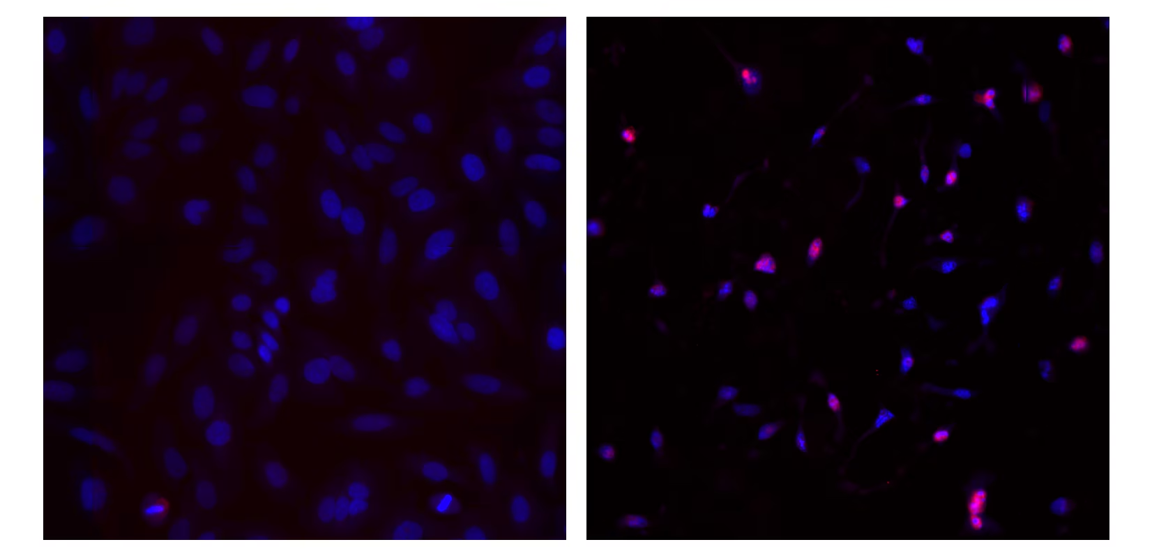
Immunofluorescent staining of human cell lines. HeLa cells were cultured overnight and treated for 4 hours with 1 μM staurosporine (right panel) or untreated (left panel). Then they were fixed, permeabilized, stained overnight with Alexa Fluor® 647 Mouse anti-Cleaved PARP (Asp 214, Cat. No. 558710) (pseudo-colored red), and counter-stained with Hoechst 33342 solution (Cat. No. 561908) (pseudo-colored blue) according to the Recommended Assay Procedure for Bioimaging. The red cleaved PARP appears pink when it is co-localized with the blue Hoechst 33342 staining. The images were captured on a BD Pathway™ 855 Bioimager System with a 20x objective and merged using BD Attovision™ software. The antibody also stains A549, and U-2 OX cells, and it works with either BD Phosflow™ Perm Buffer III or BD Perm/Wash™ buffer permeabilization (see Recommended Assay Procedure for detailed Bioimaging protocol).
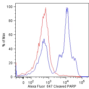
Flow cytometric analysis of cleaved PARP in camptothecin-treated Jurkat cells. Jurkat cells were either untreated (red) or treated with 5 µM camptothecin (blue) for 18 hours, and fixed, permeabilized according to the Recommended Assay Procedure for Flow Cytometry. Histograms were derived from gated events based on light scattering characteristics for Jurkat cells. Flow cytometry was performed on a BD™ LSR II flow cytometry system.


 全部商品分类


 下载产品说明书
 用小程序,查商品更便捷


 收藏
收藏
 对比
对比 咨询
咨询