


 全部商品分类


PathScan ® Phospho-DDR1 (panTyr) Sandwich ELISA Kit

 下载产品说明书 下载COA 下载SDS
 用小程序,查商品更便捷


 收藏
收藏
 对比
对比 咨询
咨询The PathScan® Phospho-DDR1 (panTyr) Sandwich ELISA Kit is a solid phase sandwich enzyme-linked immunosorbent assay (ELISA) that detects endogenous levels of tyrosine-phosphorylated DDR1 protein. A DDR1 rabbit antibody has been coated on the microwells. After incubation with cell lysates, DDR1 protein (phospho and nonphospho) is captured by the coated antibody. Following extensive washing, a phospho-tyrosine mouse mAb is added to detect captured tyrosine-phosphorylated DDR1 protein. Anti-mouse IgG, HRP-linked antibody is then used to recognize the bound detection antibody. HRP substrate TMB is added to develop color. The magnitude of the absorbance for this developed color is proportional to the quantity of DDR1 protein phosphorylated on tyrosine residues.
*Antibodies in this kit are custom formulations specific to kit.

Specificity/Sensitivity
Species Reactivity:
Human
参考图片
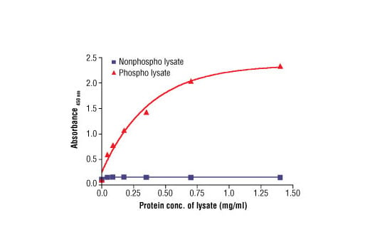
Figure 2: The relationship between protein concentration of phospho or nonphospho lysates and the absorbance at 450 nm is shown. Calu-3 cells were cultured (85% confluence) and lysed with or without the addition of phosphatase inhibitor to the lysis buffer (phospho or nonphospho lysate, respectively).
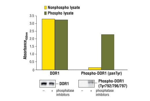
Figure 1: Constitutive phosphorylation of DDR1 in Calu-3 cells lysed in the presence of phosphatase inhibitors* (phospho lysate) is detected by PathScan® Phospho-DDR1 (panTyr) Sandwich ELISA Kit #7863 (upper, right). In contrast, a low level of phospho-DDR1 protein is detected in Calu-3 cells lysed in the absence of phosphatase inhibitors* (nonphospho lysate). Similar levels of total DDR1 protein from both nonphospho and phospho lysates are detected by PathScan® Total DDR1 Sandwich ELISA Kit #7845 (upper, left). Absorbance at 450 nm is shown in the top figure while corresponding western blots using a Phospho-DDR1 (Tyr792/796/797) rabbit antibody (right) or a total DDR1 rabbit antibody (left) are shown in the bottom figure. *Phosphatase inhibitors include sodium pyrophosphate, β-glycerophosphate and Na3VO4.