


 全部商品分类


PCNA (D3H8P) XP ® Rabbit mAb


 下载产品说明书 下载COA 下载SDS
 用小程序,查商品更便捷


 收藏
收藏
 对比
对比 咨询
咨询
Monoclonal antibody is produced by immunizing animals with a synthetic peptide corresponding to residues near the carboxy terminus of human PCNA protein.
Product Usage Information
| Application | Dilution |
|---|---|
| Western Blotting | 1:1000 |
| Immunoprecipitation | 1:50 |
| Immunohistochemistry (Paraffin) | 1:4000 - 1:16000 |
| Immunofluorescence (Frozen) | 1:400 - 1:1600 |
| Immunofluorescence (Immunocytochemistry) | 1:400 - 1:1600 |
| Flow Cytometry (Fixed/Permeabilized) | 1:100 - 1:400 |
Specificity/Sensitivity
Species Reactivity:
Human, Mouse, Rat, Monkey
Supplied in 10 mM sodium HEPES (pH 7.5), 150 mM NaCl, 100 µg/ml BSA, 50% glycerol and less than 0.02% sodium azide. Store at –20°C. Do not aliquot the antibody.
For a carrier free (BSA and azide free) version of this product see product #71395.
参考图片
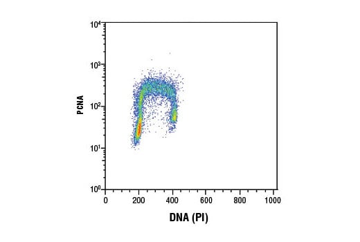
Flow cytometric analysis of Jurkat cells using PCNA (D3H8P) XP® Rabbit mAb and Propidium Iodide (PI)/RNase Staining Solution #4087 to measure DNA content. Anti-rabbit IgG (H+L), F(ab')2 Fragment (Alexa Fluor® 488 Conjugate) #4412 was used as a secondary antibody.
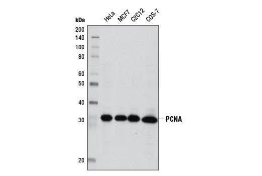
Western blot analysis of extracts from various cell lines using PCNA (D3H8P) XP® Rabbit mAb.
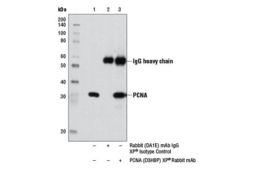
Immunoprecipitation of PCNA from HeLa cell extracts using Rabbit (DA1E) mAb IgG XP® Isotype Control #3900 (lane 2) or PCNA (D3H8P) XP® Rabbit mAb (lane 3). Lane 1 is 10% input. Western blot was performed using PCNA (D3H8P) XP® Rabbit mAb.
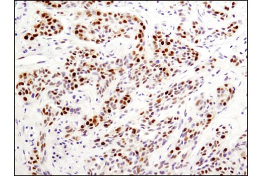
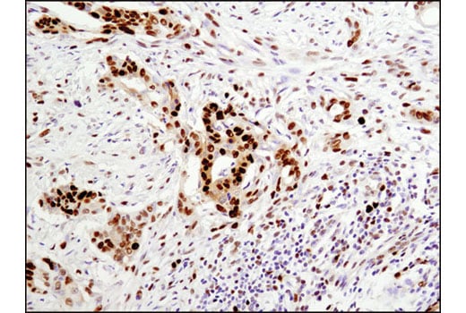
Immunohistochemical analysis of paraffin-embedded human breast carcinoma using PCNA (D3H8P) XP® Rabbit mAb.
Immunohistochemical analysis of paraffin-embedded human colon carcinoma using PCNA (D3H8P) XP® Rabbit mAb.
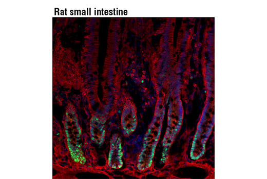
Confocal immunofluorescent analysis of rat small intestine using PCNA (D3H8P) XP® Rabbit mAb (green) showing staining of proliferative cells in intestinal crypts and β-Catenin (L54E2) Mouse mAb (IF Preferred) #2677 (red). Blue pseudocolor = DRAQ5® #4084 (fluorescent DNA dye).
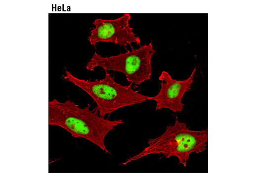
Confocal immunofluorescent analysis of HeLa cells using PCNA (D3H8P) XP® Rabbit mAb (green) and β-Actin (8H10D10) Mouse mAb #3700 (red).