


 全部商品分类


BD Horizon™ BV650 Mouse Anti-Human CD279 (PD-1)


 下载产品说明书 下载SDS
 用小程序,查商品更便捷


 收藏
收藏
 对比
对比 咨询
咨询

参考图片
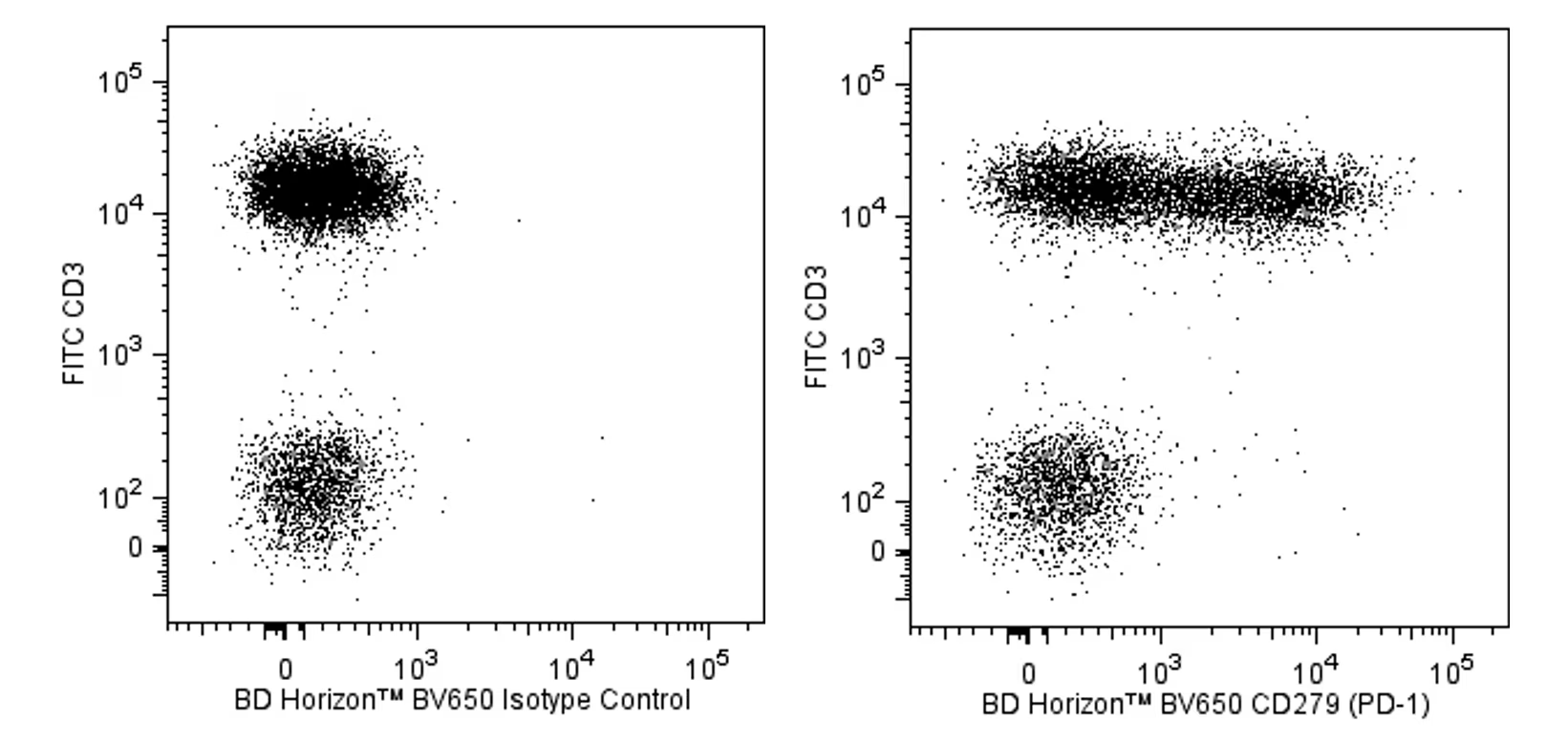
Two-color flow cytometric analysis of CD279 expression on human peripheral blood lymphocytes. Human whole blood was stained with FITC Mouse Anti-Human CD3 antibody (Cat. No. 555332/561806/561807) and either BD Horizon™ BV650 Mouse IgG1, k Isotype Control (Cat. No. 563231; Left Panel) or BD Horizon BV650 Mouse Anti-Human CD279 (PD-1) antibody (Cat. No. 564104; Right Panel). The erythrocytes were lysed with BD FACS™ Lysing Solution (Cat. No. 349202). The two-color flow cytometric dot plots show the correlated expression of CD279 (or Ig Isotype control staining) versus CD3 for gated events with the forward and side light-scatter characteristics of intact lymphocytes. Flow cytometric analysis was performed using a BD LSRFortessa™ Cell Analyzer System.
Two-color flow cytometric analysis of CD279 expression on human peripheral blood lymphocytes. Human whole blood was stained with FITC Mouse Anti-Human CD3 antibody (Cat. No. 555332/561806/561807) and either BD Horizon™ BV650 Mouse IgG1, k Isotype Control (Cat. No. 563231; Left Panel) or BD Horizon BV650 Mouse Anti-Human CD279 (PD-1) antibody (Cat. No. 564104; Right Panel). The erythrocytes were lysed with BD FACS™ Lysing Solution (Cat. No. 349202). The two-color flow cytometric dot plots show the correlated expression of CD279 (or Ig Isotype control staining) versus CD3 for gated events with the forward and side light-scatter characteristics of intact lymphocytes. Flow cytometric analysis was performed using a BD LSRFortessa™ Cell Analyzer System.