


 全部商品分类
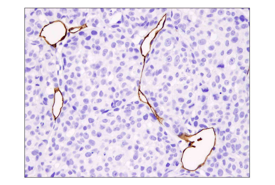


 用小程序,查商品更便捷


Monoclonal antibody is produced by immunizing animals with a synthetic peptide corresponding to residues surrounding Ala451 of mouse CD31 (PECAM-1) protein.
Product Usage Information
| Application | Dilution |
|---|---|
| Western Blotting | 1:1000 |
| Simple Western™ | 1:10 - 1:50 |
| IHC Leica Bond | 1:50 - 1:200 |
| Immunohistochemistry (Paraffin) | 1:50 - 1:200 |
Specificity/Sensitivity
Species Reactivity:
Mouse
Supplied in 10 mM sodium HEPES (pH 7.5), 150 mM NaCl, 100 µg/ml BSA, 50% glycerol and less than 0.02% sodium azide. Store at –20°C. Do not aliquot the antibody.
For a carrier free (BSA and azide free) version of this product see product #92841.
参考图片
Immunohistochemical analysis of paraffin-embedded A2058 xenograft using CD31 (PECAM-1) (D8V9E) XP® Rabbit mAb.
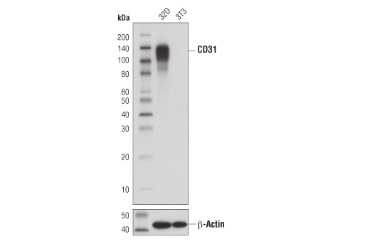
Western blot analysis of extracts from 32D (CD31/PECAM-1 positive) and 3T3 (CD31/PECAM-1 negative) cells using CD31 (PECAM-1) (D8V9E) XP® Rabbit mAb (upper) and β-Actin (D6A8) Rabbit mAb #8457 (lower).
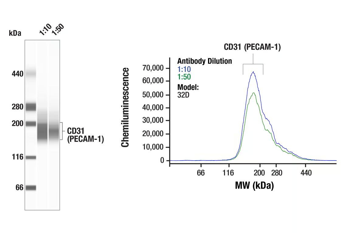
Simple Western™️ analysis of lysates (0.1mg/mL) from 32D cells using CD31 (PECAM-1) (D8V9E) Rabbit mAb #77699. The virtual lane view (left) shows the target band (as indicated) at 1:10 and 1:50 dilutions of primary antibody. The corresponding electropherogram view (right) plots chemiluminescence by molecular weight along the capillary at 1:10 (blue line) and 1:50 (green line) dilutions of primary antibody. This experiment was performed under reducing conditions on the Jess™️ Simple Western instrument from ProteinSimple, a BioTechne brand, using the 66 – 440 kDa separation module.
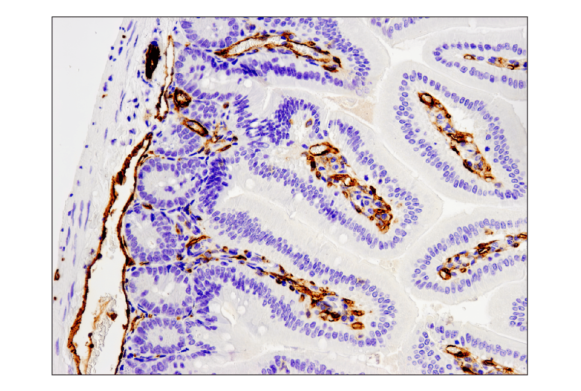
Immunohistochemical analysis of paraffin-embedded mouse small intestine using CD31 (PECAM-1) (D8V9E) XP® Rabbit mAb performed on the Leica® BOND™ Rx.
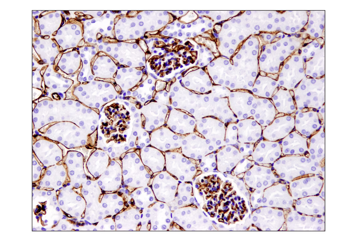
Immunohistochemical analysis of paraffin-embedded mouse kidney using CD31 (PECAM-1) (D8V9E) XP® Rabbit mAb.
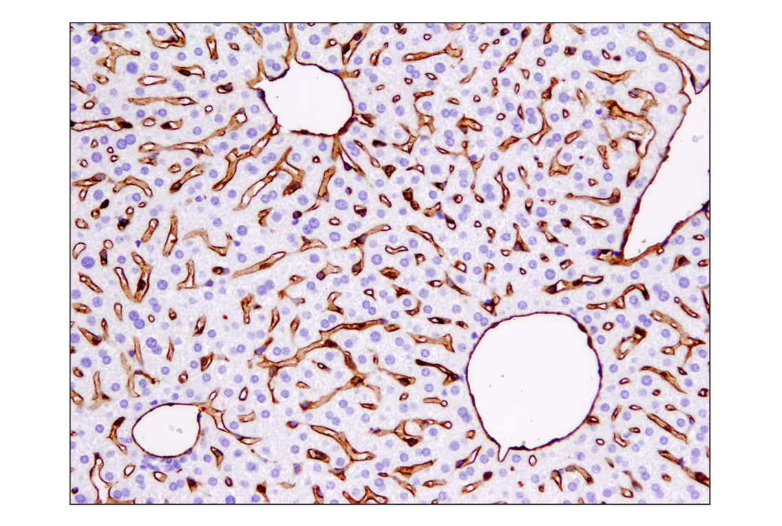
Immunohistochemical analysis of paraffin-embedded mouse liver using CD31 (PECAM-1) (D8V9E) XP® Rabbit mAb.