

 全部商品分类
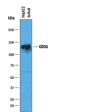


 下载产品说明书 下载SDS
 用小程序,查商品更便捷


 收藏
收藏
 对比
对比 咨询
咨询Simple Western(10 µg/mL)
Immunocytochemistry(5-15 µg/mL)

Extracellular domain
Scientific Data
 View Larger
Detection of Human CD31/PECAM‑1 by Western Blot. Western blot shows lysates of HepG2 human hepatocellular carcinoma cell line. PVDF membrane was probed with 1 µg/mL of Sheep Anti-Human CD31/PECAM-1 Antigen Affinity-purified Polyclonal Antibody (Catalog # AF806) followed by HRP-conjugated Anti-Sheep IgG Secondary Antibody (HAF016). A specific band was detected for CD31/PECAM-1 at approximately 130 kDa (as indicated). This experiment was conducted under reducing conditions and using Immunoblot Buffer Group 1.
.") View Larger
View LargerCD31/PECAM‑1 in HUVECs. CD31/PECAM-1 was detected in immersion fixed HUVEC human umbilical vein endothelial cells using 10 µg/mL Sheep Anti-Human CD31/PECAM-1 Antigen Affinity-purified Polyclonal Antibody (Catalog # AF806) for 3 hours at room temperature. Cells were stained with the NorthernLights™ 557-conjugated Anti-Sheep IgG Secondary Antibody (red; Catalog # NL010) and counterstained with DAPI (blue). View our protocol for Fluorescent ICC Staining of Cells on Coverslips.
 View Larger
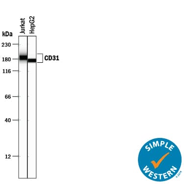
Detection of Human CD31/PECAM‑1 by Simple WesternTM. Simple Western lane view shows lysates of Jurkat human acute T cell leukemia cell line and HepG2 human hepatocellular carcinoma cell line, loaded at 0.2 mg/mL. A specific band was detected for CD31/PECAM-1 at approximately 173-186 kDa (as indicated) using 10 µg/mL of Sheep Anti-Human CD31/PECAM-1 Antigen Affinity-purified Polyclonal Antibody (Catalog # AF806) followed by 1:50 dilution of HRP-conjugated Anti-Sheep IgG Secondary Antibody (HAF016). This experiment was conducted under reducing conditions and using the 12-230 kDa separation system.
 View Larger
CD31/PECAM‑1 Specificity is Shown by Immunocytochemistry in Knockout Cell Line. CD31/PECAM‑1 was detected in immersion fixed THP‑1 human acute monocytic leukemia cell line but is not detected in CD31/PECAM‑1 knockout (KO) THP‑1 human cell line using Sheep Anti-Human CD31/PECAM‑1 Antigen Affinity-purified Polyclonal Antibody (Catalog # AF806) at 5 µg/mL for 3 hours at room temperature. Cells were stained using the NorthernLights™ 557-conjugated Anti-Sheep IgG Secondary Antibody (red; NL010) and counterstained with DAPI (blue). Specific staining was localized to cell membrane. Staining was performed using our protocol for Fluorescent ICC Staining of Non-adherent Cells.
 View Larger
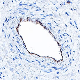
Detection of CD31/PECAM‑1 in human liver. CD31/PECAM‑1 was detected in immersion fixed paraffin-embedded sections of human liver using Sheep Anti-Human CD31/PECAM‑1 Antigen Affinity-purified Polyclonal Antibody (Catalog # AF806) at 5 µg/mL for 1 hour at room temperature followed by incubation with the Anti-Goat IgG VisUCyte™ HRP Polymer Antibody (Catalog # VC004). Before incubation with the primary antibody, tissue was subjected to heat-induced epitope retrieval using VisUCyte Antigen Retrieval Reagent-Basic (Catalog # VCTS021). Tissue was stained using DAB (brown) and counterstained with hematoxylin (blue). Specific staining was localized to endothelial cells. View our protocol for IHC Staining with VisUCyte HRP Polymer Detection Reagents.
 View Larger
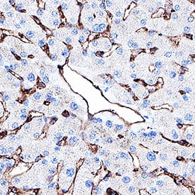
Detection of CD31/PECAM‑1 in human liver. CD31/PECAM‑1 was detected in immersion fixed paraffin-embedded sections of human liver using Sheep Anti-Human CD31/PECAM‑1 Antigen Affinity-purified Polyclonal Antibody (Catalog # AF806) at 5 µg/mL for 1 hour at room temperature followed by incubation with the Anti-Goat IgG VisUCyte™ HRP Polymer Antibody (Catalog # VC004). Before incubation with the primary antibody, tissue was subjected to heat-induced epitope retrieval using VisUCyte Antigen Retrieval Reagent-Basic (Catalog # VCTS021). Tissue was stained using DAB (brown) and counterstained with hematoxylin (blue). Specific staining was localized to endothelial cells and sinusoids. View our protocol for IHC Staining with VisUCyte HRP Polymer Detection Reagents.
 View Larger
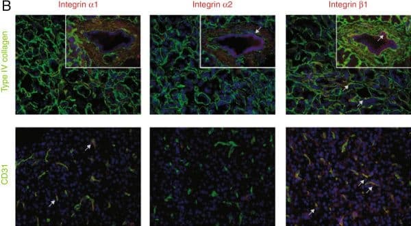
Detection of Human CD31/PECAM-1 by Immunocytochemistry/Immunofluorescence Expression pattern of integrin receptors and type IV collagen in normal pancreatic tissue. A. H&E staining of normal exocrine pancreas with acini structures and a duct in the insert. The area shown is representative for all panels in B. B. Merged immunofluorescence staining of integrin alpha 1, alpha 2, and beta 1 (in red), and type IV collagen (green in upper row), or the endothelial marker CD31 (green in lower row). Type IV collagen is present in the vascular-, ductal-, and acini-BMs. Integrin alpha 1 is only expressed in endothelial cells and integrin alpha 2 only in the ductal epithelium. Integrin beta 1 is found in the ductal-, endothelial-, and at the base of the acini-cells. Arrows indicate examples of colocalization. Magnification x40 in all panels. Cell nuclei are stained by DAPI (in blue). Image collected and cropped by CiteAb from the following publication (https://bmccancer.biomedcentral.com/articles/10.1186/1471-2407-13-154), licensed under a CC-BY license. Not internally tested by R&D Systems.
Human CD31/PECAM-1 Antibody Summary
Extracellular domain
Applications
Please Note: Optimal dilutions should be determined by each laboratory for each application. General Protocols are available in the Technical Information section on our website.
Simple Western(10 µg/mL)
Immunocytochemistry(5-15 µg/mL)
Background: CD31/PECAM-1
The CD31 adhesion molecule, also known as PECAM-1, is expressed in large amounts on endothelial cells at intercellular junctions and on T cell subsets, and to a lesser extent on platelets and most other leukocytes such as monocytes and neutrophils. CD31 binds to itself homotypically, and also to the leukocyte integrin alpha v beta 3 heterotypically. CD31 is required for the transendothelial migration of leukocytes through intercellular junctions of vascular endothelial cells. CD31 has been found in human plasma, and the presence of this circulating isoform is suggested to modulate the transendothelial migration of leukocytes.
Preparation and Storage
- 12 months from date of receipt, -20 to -70 °C as supplied.
- 1 month, 2 to 8 °C under sterile conditions after reconstitution.
- 6 months, -20 to -70 °C under sterile conditions after reconstitution.
参考图片
Detection of Human CD31/PECAM‑1 by Western Blot. Western blot shows lysates of HepG2 human hepatocellular carcinoma cell line. PVDF membrane was probed with 1 µg/mL of Sheep Anti-Human CD31/PECAM‑1 Antigen Affinity-purified Polyclonal Antibody (Catalog # AF806) followed by HRP-conjugated Anti-Goat IgG Secondary Antibody (Catalog # HAF019). A specific band was detected for CD31/PECAM‑1 at approximately 130 kDa (as indicated). This experiment was conducted under reducing conditions and using Immunoblot Buffer Group 1.
CD31/PECAM‑1 in HUVECs. CD31/PECAM‑1 was detected in immersion fixed HUVEC human umbilical vein endothelial cells using 10 µg/mL Sheep Anti-Human CD31/PECAM‑1 Antigen Affinity-purified Polyclonal Antibody (Catalog # AF806) for 3 hours at room temperature. Cells were stained with the NorthernLights™ 557-conjugated Anti-Sheep IgG Secondary Antibody (red; Catalog # NL010) and counterstained with DAPI (blue). View our protocol for Fluorescent ICC Staining of Cells on Coverslips.
Detection of Human CD31/PECAM‑1 by Simple WesternTM. Simple Western lane view shows lysates of Jurkat human acute T cell leukemia cell line and HepG2 human hepatocellular carcinoma cell line, loaded at 0.2 mg/mL. A specific band was detected for CD31/PECAM‑1 at approximately
173-186 kDa (as indicated) using 10 µg/mL of Sheep Anti-Human CD31/PECAM‑1 Antigen Affinity-purified Polyclonal Antibody (Catalog # AF806) followed by 1:50 dilution of HRP-conjugated Anti-Sheep IgG Secondary Antibody (Catalog # HAF016). This experiment was conducted under reducing conditions and using the
12-230 kDa separation system.